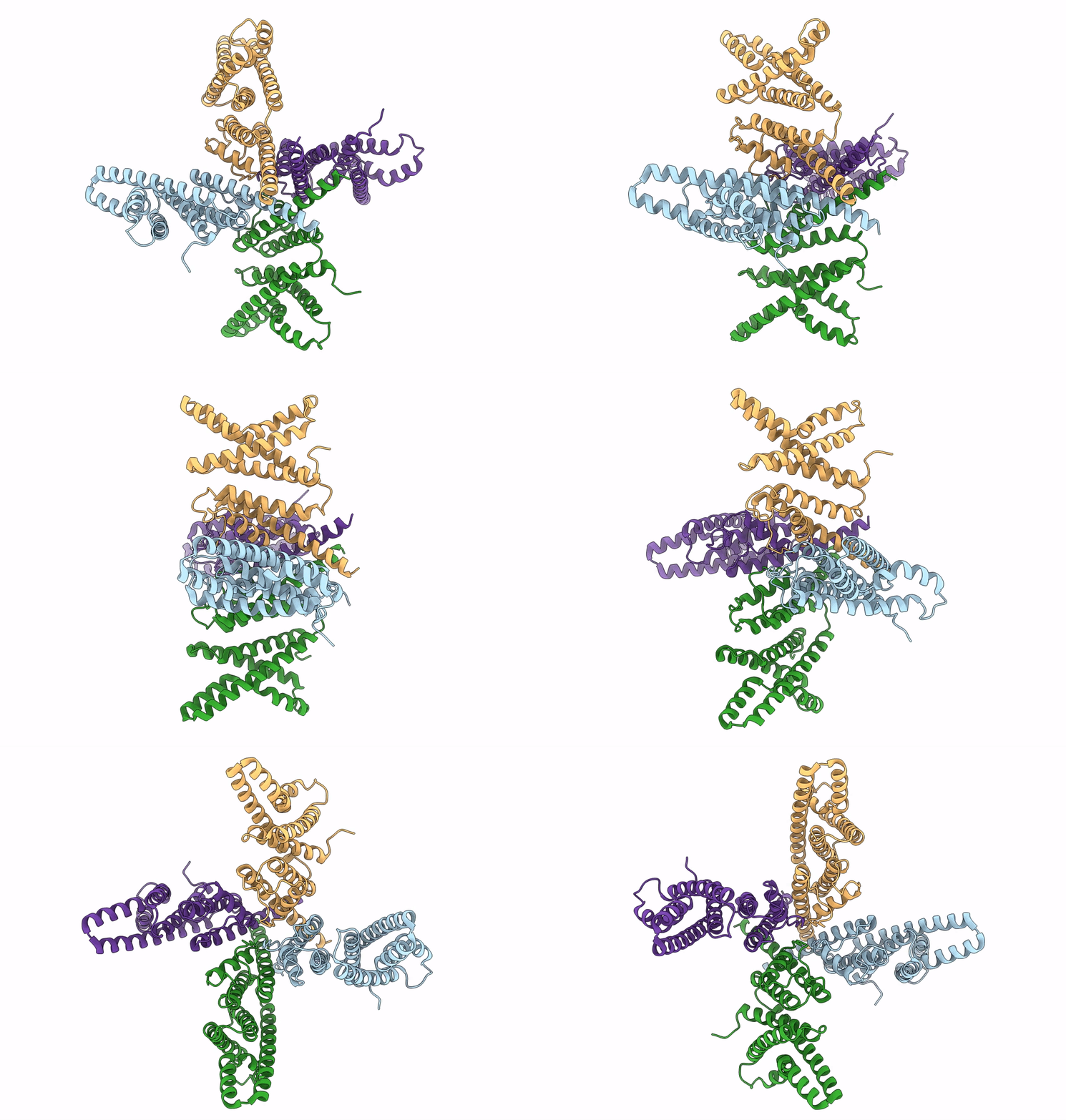
6UWM image
Deposition Date 2019-11-05
Release Date 2019-12-04
Last Version Date 2024-03-06
Entry Detail
PDB ID:
6UWM
Title:
Single particle cryo-EM structure of KvAP
Biological Source:
Source Organism:
Method Details:
Experimental Method:
Resolution:
5.90 Å
Aggregation State:
PARTICLE
Reconstruction Method:
SINGLE PARTICLE
Macromolecular Entities
Structures with similar UniProt ID
Protein Blast
Polymer Type:polypeptide(L)
Molecule:Voltage-gated potassium channel
Gene (Uniprot):APE_0955
Chain IDs:A, B (auth: C), C (auth: B), D
Chain Length:287
Number of Molecules:4
Biological Source:Aeropyrum pernix
Ligand Molecules
Primary Citation
Cryo-EM structure of the KvAP channel reveals a non-domain-swapped voltage sensor topology.
Elife 8 ? ? (2019)
PMID: 31755864 DOI: 10.7554/eLife.52164

Abstact

Conductance in voltage-gated ion channels is regulated by membrane voltage through structural domains known as voltage sensors. A single structural class of voltage sensor domain exists, but two different modes of voltage sensor attachment to the pore occur in nature: domain-swapped and non-domain-swapped. Since the more thoroughly studied Kv1-7, Nav and Cav channels have domain-swapped voltage sensors, much less is known about non-domain-swapped voltage-gated ion channels. In this paper, using cryo-EM, we show that KvAP from Aeropyrum pernix has non-domain-swapped voltage sensors as well as other unusual features. The new structure, together with previous functional data, suggests that KvAP and the Shaker channel, to which KvAP is most often compared, probably undergo rather different voltage-dependent conformational changes when they open.

Legend

Protein

Chemical

Disease

Primary Citation of related structures
Feedback Form
Name
Email
Institute
Feedback