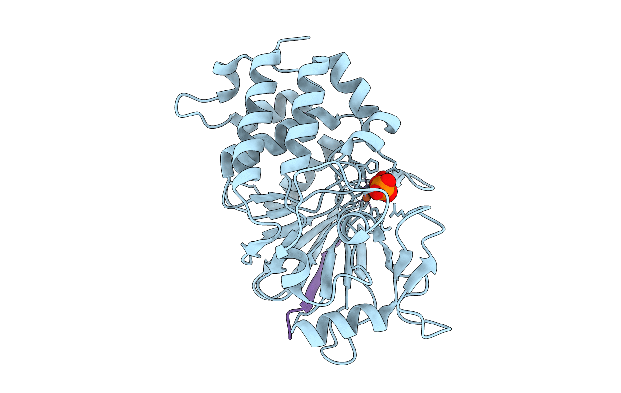
6UUQ image
Entry Detail
PDB ID:
6UUQ
Title:
Structure of Calcineurin bound to RCAN1
Biological Source:
Source Organism:
Host Organism:
PDB Version:
Deposition Date:
2019-10-31
Release Date:
2020-09-09
Method Details:
Experimental Method:
Resolution:
1.85 Å
R-Value Free:
0.20
R-Value Work:
0.16
R-Value Observed:
0.16
Space Group:
P 21 21 21
Macromolecular Entities
Polymer Type:polypeptide(L)
Description:Serine/threonine-protein phosphatase 2B catalytic subunit alpha isoform
Chain IDs:A
Chain Length:316
Number of Molecules:1
Biological Source:Homo sapiens
Polymer Type:polypeptide(L)
Description:Calcipressin-1
Mutations:V158I, H159T
Chain IDs:B
Chain Length:40
Number of Molecules:1
Biological Source:Homo sapiens
Primary Citation
The structure of the RCAN1:CN complex explains the inhibition of and substrate recruitment by calcineurin.
Sci Adv 6 ? ? (2020)
PMID: 32936779 DOI: 10.1126/sciadv.aba3681

Abstact

Regulator of calcineurin 1 (RCAN1) is an endogenous inhibitor of the Ser/Thr phosphatase calcineurin (CN). It has been shown that excessive inhibition of CN is a critical factor for Down syndrome and Alzheimer's disease. Here, we determined RCAN1's mode of action. Using a combination of structural, biophysical, and biochemical studies, we show that RCAN1 inhibits CN via multiple routes: first, by blocking essential substrate recruitment sites and, second, by blocking the CN active site using two distinct mechanisms. We also show that phosphorylation either inhibits RCAN1-CN assembly or converts RCAN1 into a weak inhibitor, which can be reversed by CN via dephosphorylation. This highlights the interplay between posttranslational modifications in regulating CN activity. Last, this work advances our understanding of how active site inhibition of CN can be achieved in a highly specific manner. Together, these data provide the necessary road map for targeting multiple neurological disorders.

Legend

Protein

Chemical

Disease

Primary Citation of related structures