
Deposition Date
2019-10-25
Release Date
2020-01-22
Last Version Date
2024-10-16
Entry Detail
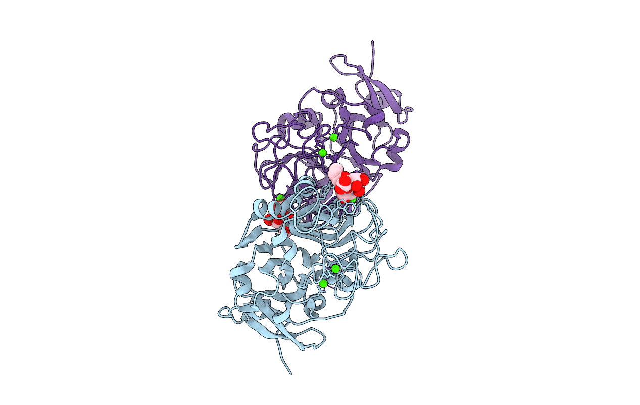
PDB ID:
6USC
Keywords:
Title:
Structure of Human Intelectin-1 in complex with KO
Biological Source:
Source Organism(s):
Homo sapiens (Taxon ID: 9606)
Expression System(s):
Method Details:
Experimental Method:
Resolution:
1.59 Å
R-Value Free:
0.18
R-Value Work:
0.15
R-Value Observed:
0.15
Space Group:
P 21 3
