
Deposition Date
2019-10-21
Release Date
2020-05-13
Last Version Date
2025-04-02
Entry Detail
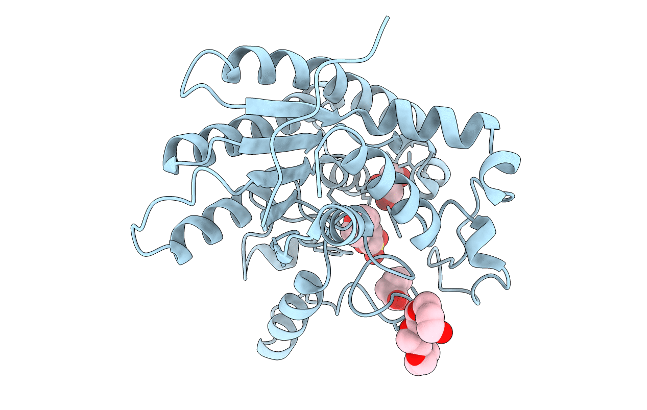
PDB ID:
6UQV
Keywords:
Title:
Crystal structure of ChoE, a bacterial acetylcholinesterase from Pseudomonas aeruginosa
Biological Source:
Source Organism:
Host Organism:
Method Details:
Experimental Method:
Resolution:
1.35 Å
R-Value Free:
0.17
R-Value Work:
0.15
R-Value Observed:
0.15
Space Group:
C 2 2 21
