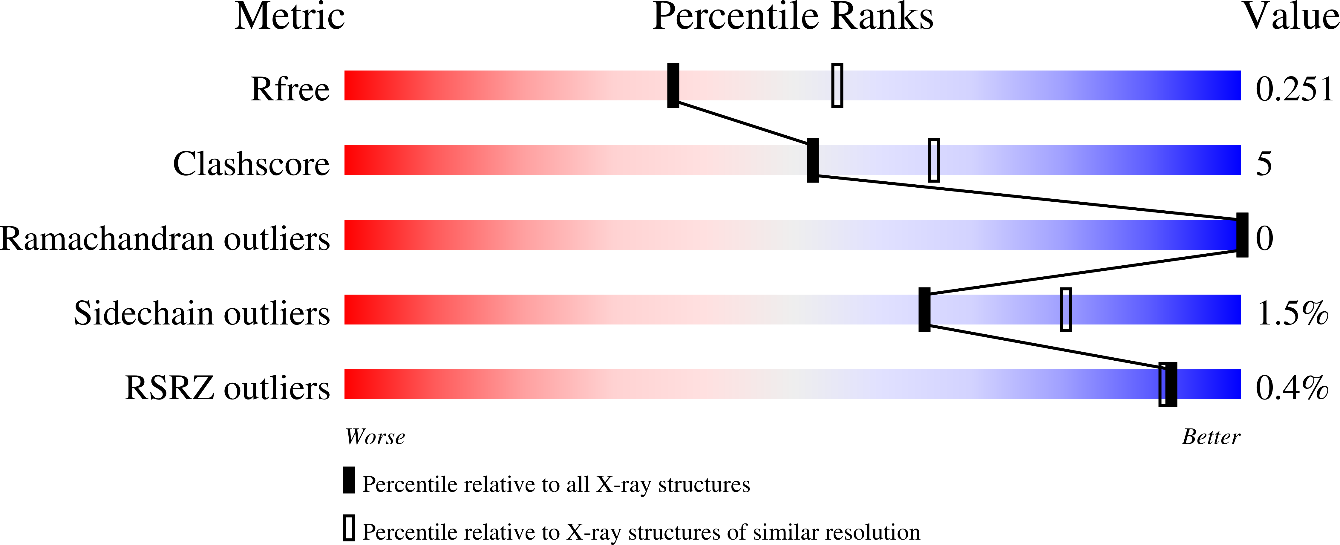
6UOD image
Deposition Date 2019-10-14
Release Date 2020-10-21
Last Version Date 2024-11-06
Entry Detail
PDB ID:
6UOD
Keywords:
Title:
Asparaginase II from Escherichia coli
Biological Source:
Source Organism:
Escherichia coli (Taxon ID: 562)
Host Organism:
Method Details:
Experimental Method:
Resolution:
2.40 Å
R-Value Free:
0.25
R-Value Work:
0.19
R-Value Observed:
0.20
Space Group:
C 1 2 1
Macromolecular Entities
Polymer Type:polypeptide(L)
Molecule:L-asparaginase 2
Chain IDs:A, B, C, D
Chain Length:326
Number of Molecules:4
Biological Source:Escherichia coli
Primary Citation
Biophysical characterization of two commercially available preparations of the drug containing Escherichia coli L-Asparaginase 2.
Biophys.Chem. 271 106554 106554 (2021)
PMID: 33607531 DOI: 10.1016/j.bpc.2021.106554

Abstact

The hydrolysis of asparagine and glutamine by L-asparaginase has been used to treat acute lymphoblastic leukemia for over four decades. Each L-asparaginase monomer has a long loop that closes over the active site upon substrate binding, acting as a lid. Here we present a comparative study of two commercially available preparations of the drug containing Escherichia coli L-Asparaginase 2 (EcA2), performed by a comprehensive array of biophysical and biochemical approaches. We report the oligomeric landscape and conformational and dynamic plasticity of E. coli type 2 L-asparaginase present in two different formulations, and its relationship with L-aspartic acid, which is present in Aginasa, but not in Leuginase. The L-Asp present in Aginasa formulation was found to provide to EcA2 a resistance to in vitro proteolysis. EcA2 shows a composition of monomers and oligomers up to tetramers, which is mostly not altered in the presence of L-Asp. Ion-mobility spectrometry-mass spectrometry reveals two conformers for the monomeric EcA2, and that monomeric species has sufficient capacity for selective binding to L-Asp and L-Glu. The N-terminal loop of the EcA2 present in Leuginase, which is part of the active site is disordered, but it gets ordered in the presence of L-Asp, while L-Glu only does so to a limited extent. These data provide new insights on the mechanistic of ligand recognition by EcA2, and the impact of formulation in its conformational diversity landscape.

Legend

Protein

Chemical

Disease

Primary Citation of related structures