
Deposition Date
2019-10-09
Release Date
2021-04-14
Last Version Date
2024-11-06
Entry Detail
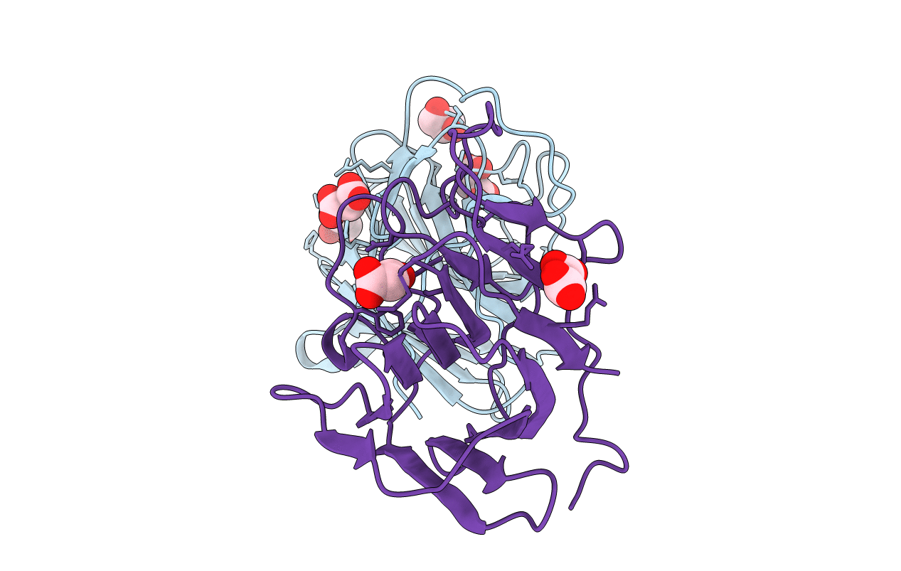
PDB ID:
6UMA
Keywords:
Title:
Crystal structure of the TRIM7 B30.2 domain at 1.6 angstrom resolution
Biological Source:
Source Organism(s):
Homo sapiens (Taxon ID: 9606)
Expression System(s):
Method Details:
Experimental Method:
Resolution:
1.60 Å
R-Value Free:
0.20
R-Value Work:
0.17
R-Value Observed:
0.17
Space Group:
P 21 21 21
