Deposition Date
2019-10-02
Release Date
2020-09-30
Last Version Date
2024-10-30
Entry Detail
PDB ID:
6UJ6
Keywords:
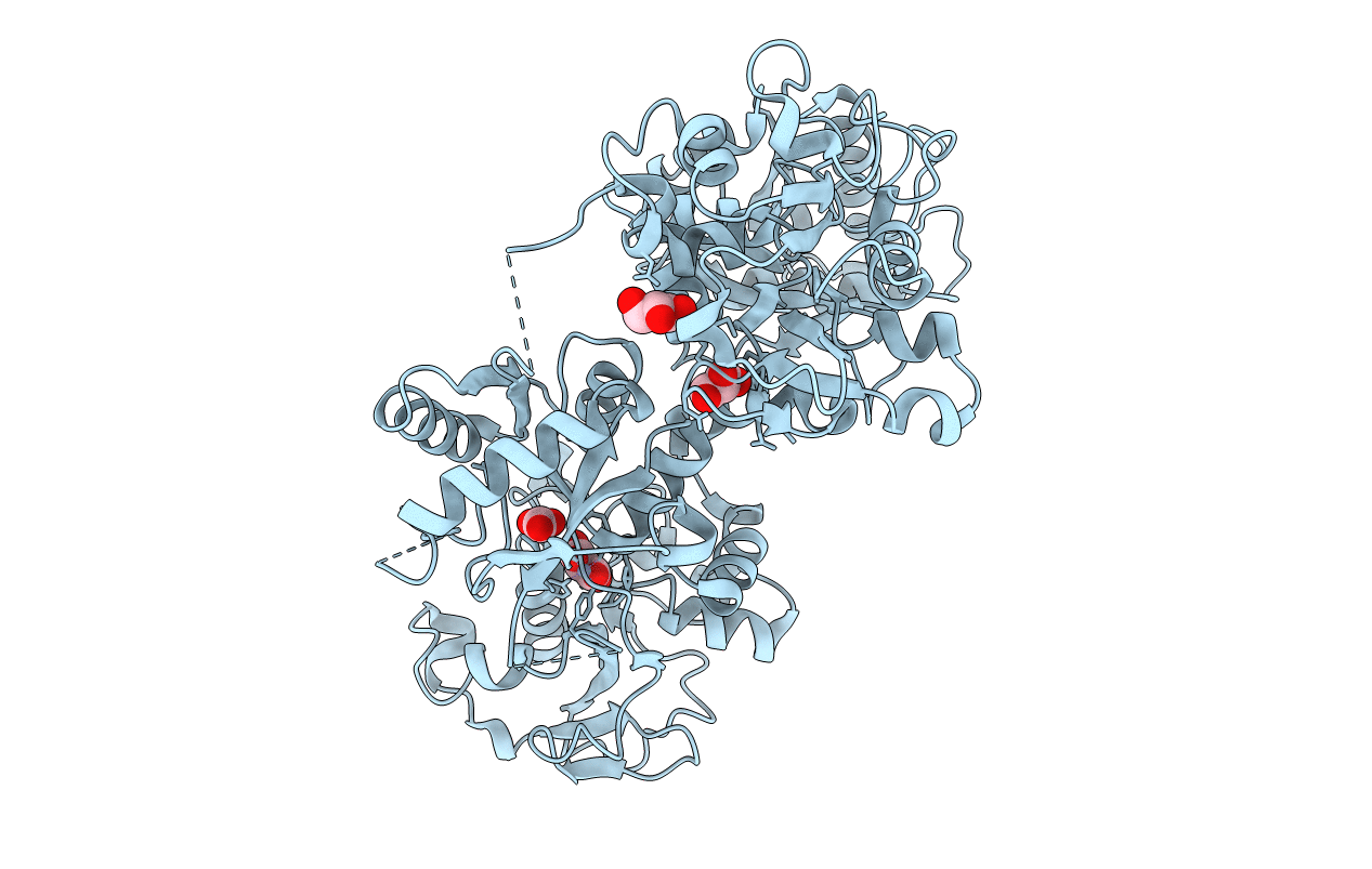
Title:
X-ray Crystal Structure of Chromium-transferrin with Synergistic Anion Malonate
Biological Source:
Source Organism(s):
Homo sapiens (Taxon ID: 9606)
Method Details:
Experimental Method:
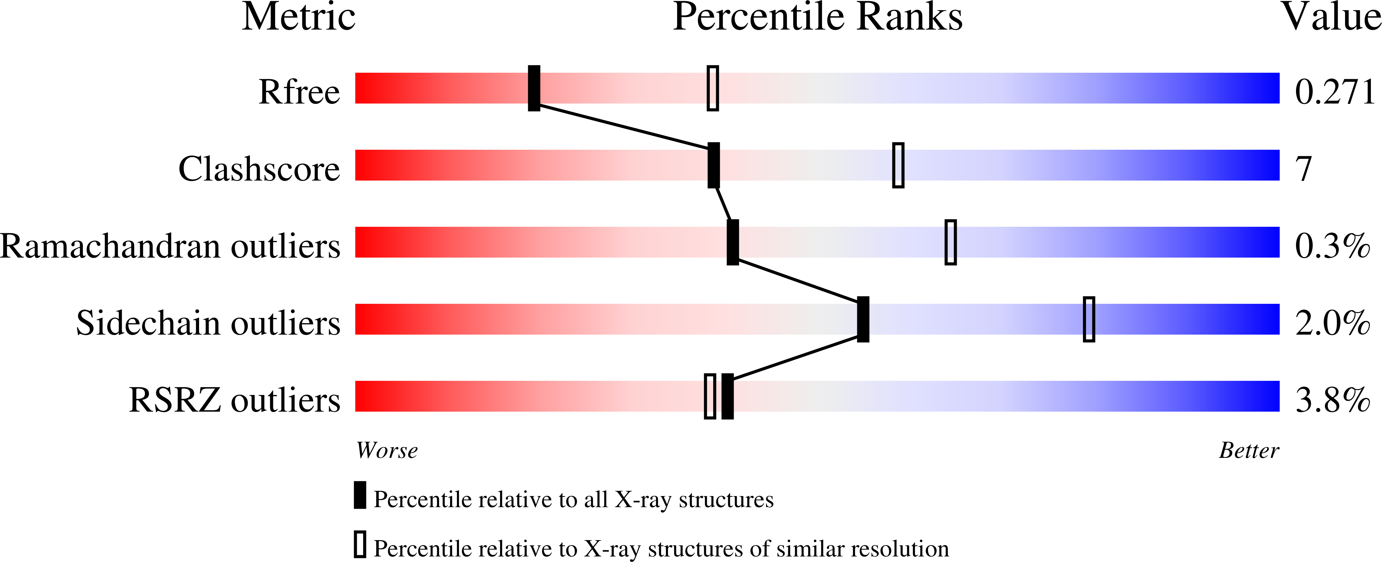
Resolution:
2.68 Å
R-Value Free:
0.27
R-Value Work:
0.22
R-Value Observed:
0.22
Space Group:
C 2 2 21