6UEX image
Deposition Date 2019-09-23
Release Date 2020-01-29
Last Version Date 2023-10-11
Entry Detail
PDB ID:
6UEX
Keywords:
Title:
Crystal structure of S. aureus LcpA in complex with octaprenyl-pyrophosphate-GlcNAc
Biological Source:
Source Organism:
Method Details:
Experimental Method:
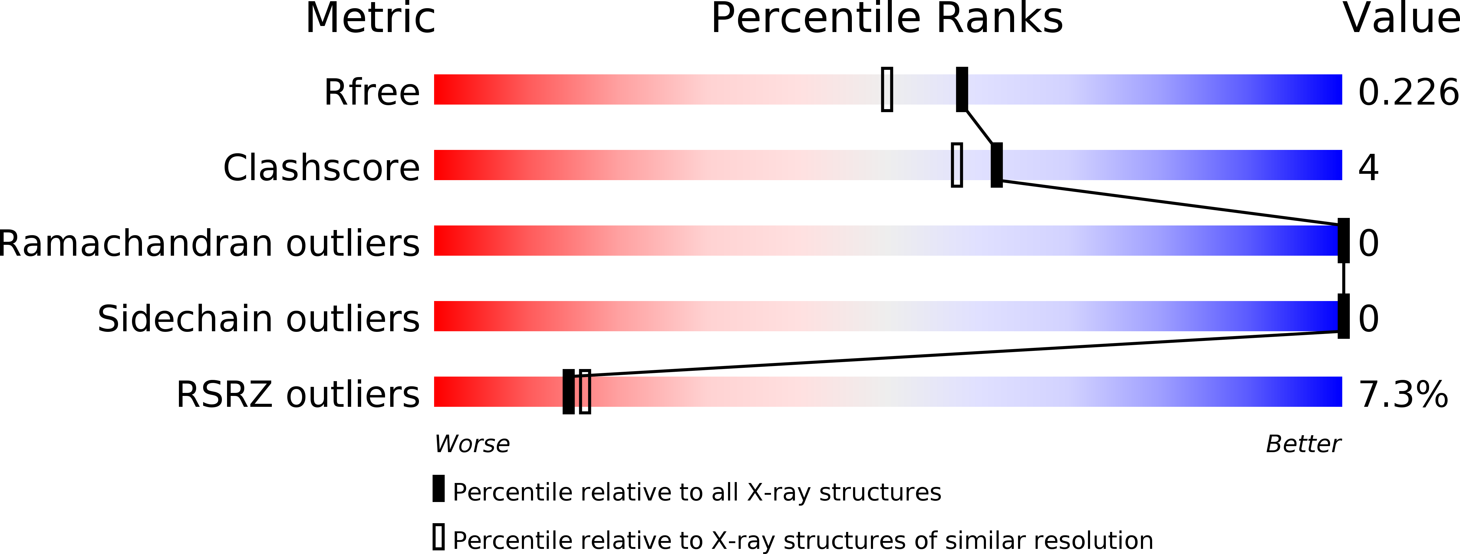
Resolution:
1.90 Å
R-Value Free:
0.22
R-Value Work:
0.19
R-Value Observed:
0.19
Space Group:
C 2 2 21
Macromolecular Entities
Polymer Type:polypeptide(L)
Molecule:Regulatory protein MsrR
Gene (Uniprot):msrR
Chain IDs:A
Chain Length:273
Number of Molecules:1
Biological Source:Staphylococcus aureus (strain N315)
Primary Citation
Crystallographic analysis ofStaphylococcus aureusLcpA, the primary wall teichoic acid ligase.
J.Biol.Chem. 295 2629 2639 (2020)
PMID: 31969390 DOI: 10.1074/jbc.RA119.011469

Abstact

Gram-positive bacteria, including major clinical pathogens such as Staphylococcus aureus, are becoming increasingly drug-resistant. Their cell walls are composed of a thick layer of peptidoglycan (PG) modified by the attachment of wall teichoic acid (WTA), an anionic glycopolymer that is linked to pathogenicity and regulation of cell division and PG synthesis. The transfer of WTA from lipid carriers to PG, catalyzed by the LytR-CpsA-Psr (LCP) enzyme family, offers a unique extracellular target for the development of new anti-infective agents. Inhibitors of LCP enzymes have the potential to manage a wide range of bacterial infections because the target enzymes are implicated in the assembly of many other bacterial cell wall polymers, including capsular polysaccharide of streptococcal species and arabinogalactan of mycobacterial species. In this study, we present the first crystal structure of S. aureus LcpA with bound substrate at 1.9 Å resolution and those of Bacillus subtilis LCP enzymes, TagT, TagU, and TagV, in the apo form at 1.6-2.8 Å resolution. The structures of these WTA transferases provide new insight into the binding of lipid-linked WTA and enable assignment of the catalytic roles of conserved active-site residues. Furthermore, we identified potential subsites for binding the saccharide core of PG using computational docking experiments, and multiangle light-scattering experiments disclosed novel oligomeric states of the LCP enzymes. The crystal structures and modeled substrate-bound complexes of the LCP enzymes reported here provide insights into key features linked to substrate binding and catalysis and may aid the structure-guided design of specific LCP inhibitors.

Legend

Protein

Chemical

Disease

Primary Citation of related structures