Deposition Date
2019-09-18
Release Date
2021-02-03
Last Version Date
2023-10-11
Entry Detail
PDB ID:
6UD5
Keywords:
Title:
Crystal structure of human tryptophan 2,3-dioxygenase in complex with carbon monoxide and tryptophan
Biological Source:
Source Organism(s):
Homo sapiens (Taxon ID: 9606)
Expression System(s):
Method Details:
Experimental Method:
Resolution:
2.05 Å
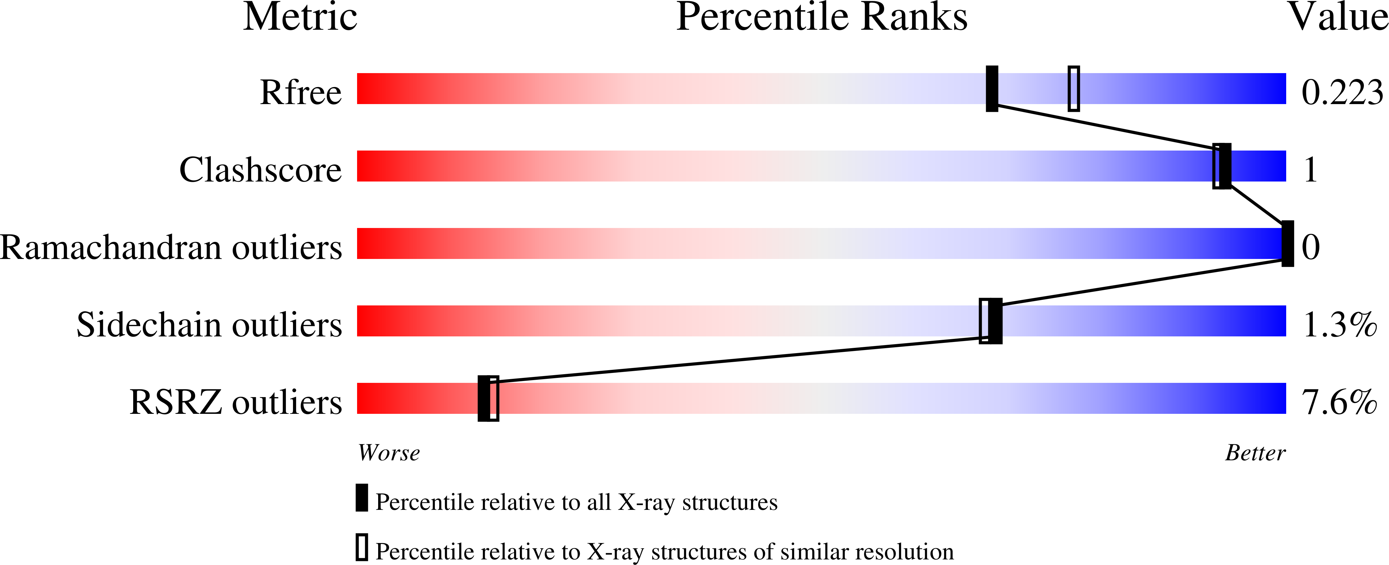
R-Value Free:
0.21
R-Value Work:
0.18
R-Value Observed:
0.18
Space Group:
P 21 21 2