Deposition Date
2019-09-11
Release Date
2020-07-29
Last Version Date
2023-10-11
Entry Detail
PDB ID:
6UBH
Keywords:
Title:
Structure of the MM7 Erbin PDZ variant in complex with a high-affinity peptide
Biological Source:
Source Organism(s):
Homo sapiens (Taxon ID: 9606)
synthetic construct (Taxon ID: 32630)
synthetic construct (Taxon ID: 32630)
Expression System(s):
Method Details:
Experimental Method:
Resolution:
1.80 Å
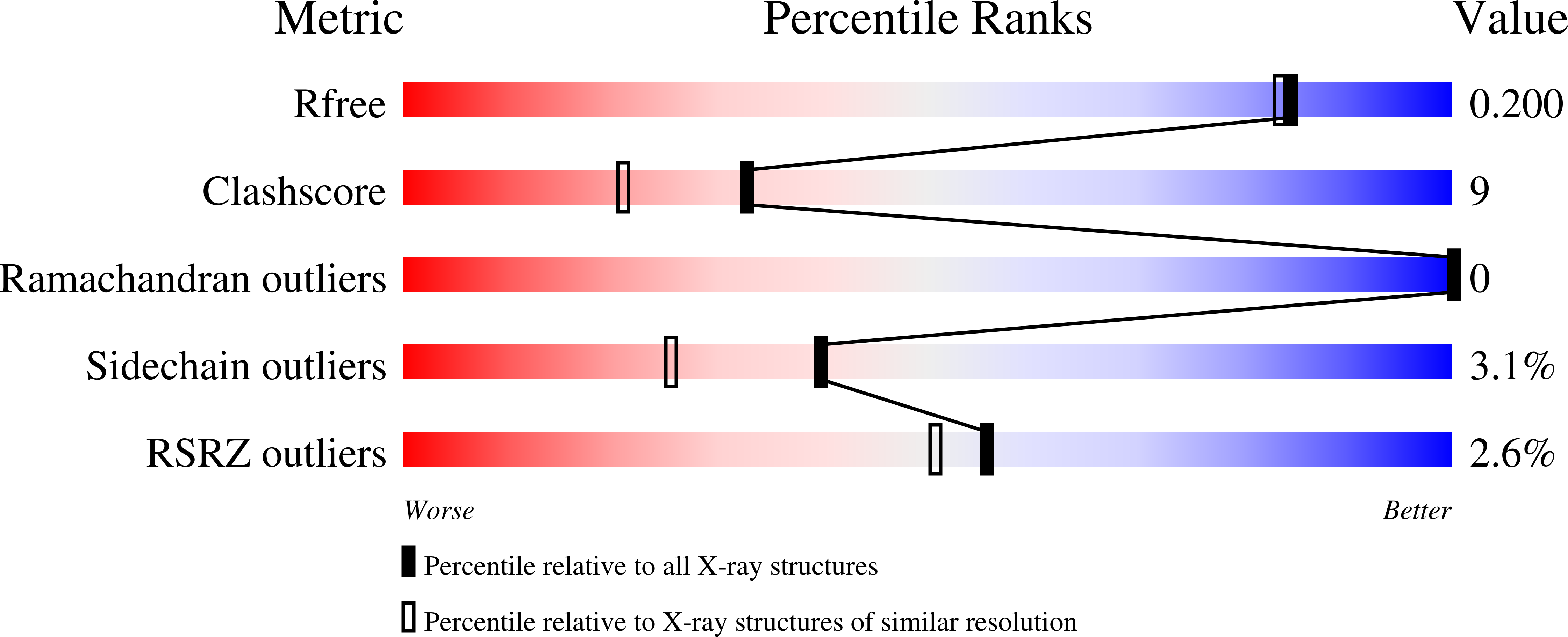
R-Value Free:
0.22
R-Value Work:
0.19
R-Value Observed:
0.21
Space Group:
P 1