6UAV image
Entry Detail
PDB ID:
6UAV
Keywords:
Title:
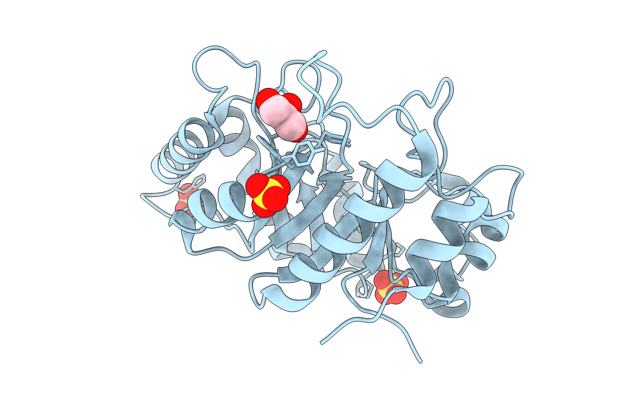
Crystal structure of a GH128 (subgroup II) endo-beta-1,3-glucanase from Pseudomonas viridiflava (PvGH128_II)
Biological Source:
Source Organism:
Host Organism:
PDB Version:
Deposition Date:
2019-09-11
Release Date:
2020-05-20
Method Details:
Experimental Method:
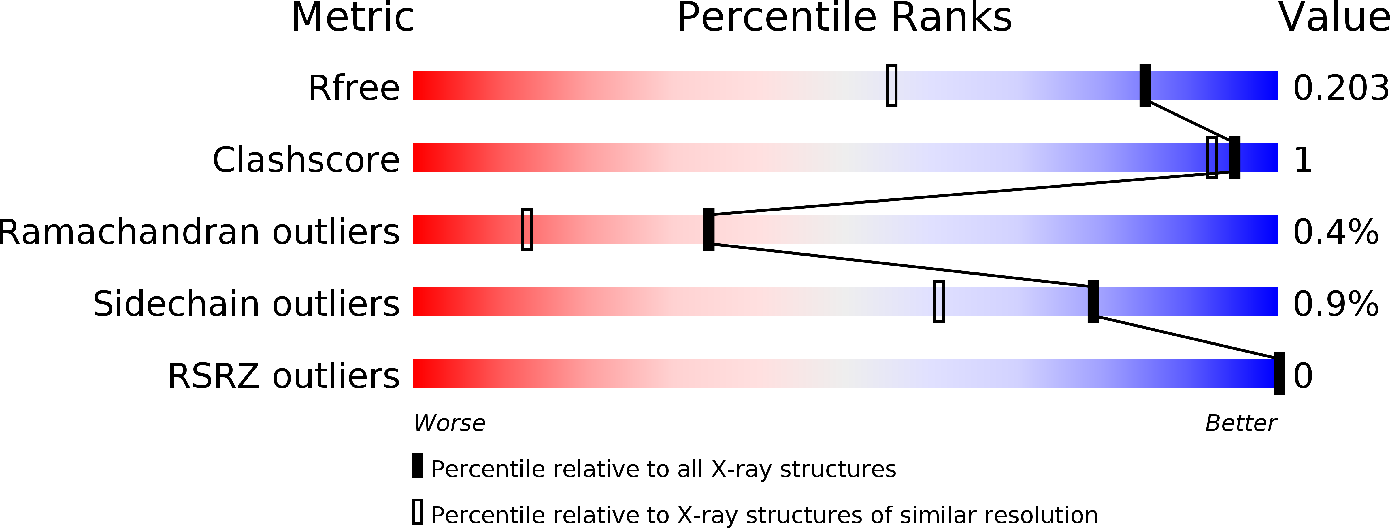
Resolution:
1.50 Å
R-Value Free:
0.20
R-Value Work:
0.17
R-Value Observed:
0.17
Space Group:
C 1 2 1
Macromolecular Entities
Polymer Type:polypeptide(L)
Description:Glyco_hydro_cc domain-containing protein
Chain IDs:A
Chain Length:259
Number of Molecules:1
Biological Source:Pseudomonas viridiflava
Primary Citation

Abstact

The fundamental and assorted roles of β-1,3-glucans in nature are underpinned on diverse chemistry and molecular structures, demanding sophisticated and intricate enzymatic systems for their processing. In this work, the selectivity and modes of action of a glycoside hydrolase family active on β-1,3-glucans were systematically investigated combining sequence similarity network, phylogeny, X-ray crystallography, enzyme kinetics, mutagenesis and molecular dynamics. This family exhibits a minimalist and versatile (α/β)-barrel scaffold, which can harbor distinguishing exo or endo modes of action, including an ancillary-binding site for the anchoring of triple-helical β-1,3-glucans. The substrate binding occurs via a hydrophobic knuckle complementary to the canonical curved conformation of β-1,3-glucans or through a substrate conformational change imposed by the active-site topology of some fungal enzymes. Together, these findings expand our understanding of the enzymatic arsenal of bacteria and fungi for the breakdown and modification of β-1,3-glucans, which can be exploited for biotechnological applications.

Legend

Protein

Chemical

Disease

Primary Citation of related structures