
Deposition Date
2019-08-15
Release Date
2020-04-22
Last Version Date
2023-11-15
Entry Detail
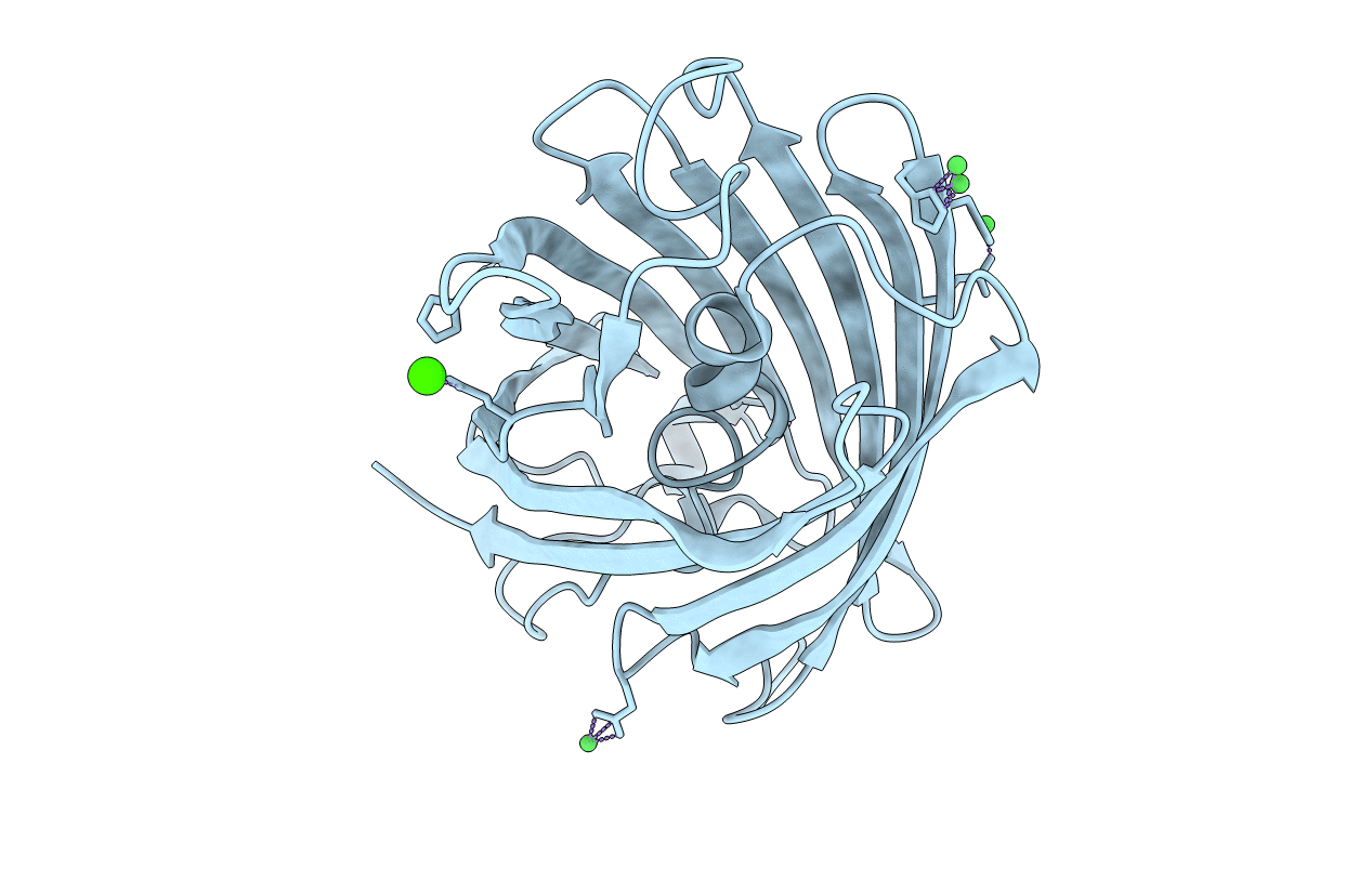
PDB ID:
6U1A
Keywords:
Title:
Crystal Structure of Fluorescent Protein FusionRed
Biological Source:
Source Organism:
Entacmaea quadricolor (Taxon ID: 6118)
Host Organism:
Method Details:
Experimental Method:
Resolution:
1.09 Å
R-Value Free:
0.14
R-Value Work:
0.12
R-Value Observed:
0.12
Space Group:
P 21 21 21
