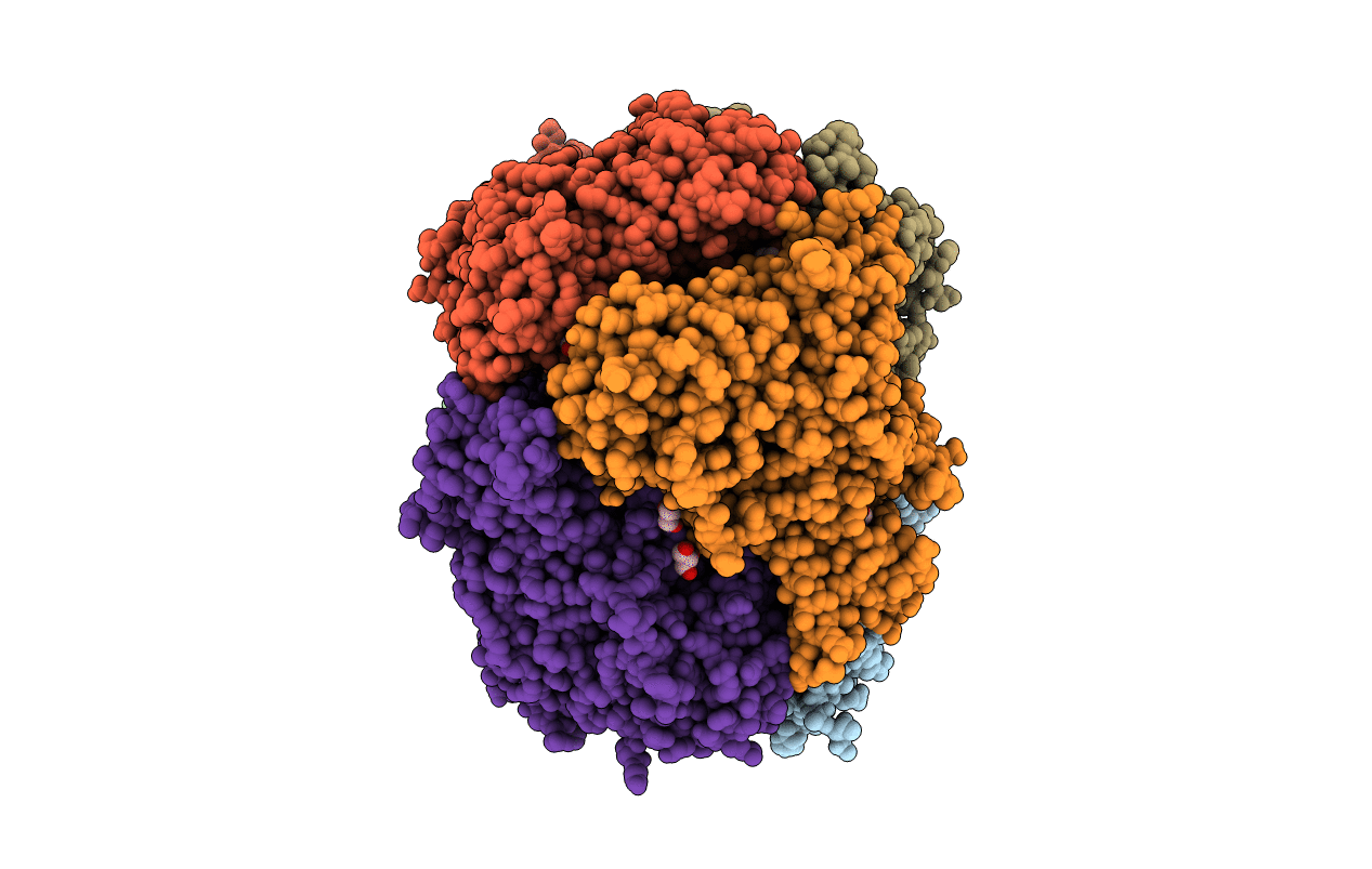
6U0P image
Entry Detail
PDB ID:
6U0P
Keywords:
Title:
Crystal structure of PieE, the flavin-dependent monooxygenase involved in the biosynthesis of piericidin A1
Biological Source:
PDB Version:
Deposition Date:
2019-08-14
Release Date:
2020-03-11
Method Details:
Experimental Method:
Resolution:
2.02 Å
R-Value Free:
0.20
R-Value Work:
0.16
R-Value Observed:
0.17
Space Group:
P 21 21 21
Macromolecular Entities
Polymer Type:polypeptide(L)
Description:2,4-dichlorophenol 6-monooxygenase
Chain IDs:A, B, C, D, E, F
Chain Length:601
Number of Molecules:6
Biological Source:Streptomyces sp. SCSIO 03032
Primary Citation
Structural analyses of the Group A flavin-dependent monooxygenase PieE reveal a sliding FAD cofactor conformation bridging OUT and IN conformations.
J.Biol.Chem. 295 4709 4722 (2020)
PMID: 32111738 DOI: 10.1074/jbc.RA119.011212

Abstact

Group A flavin-dependent monooxygenases catalyze the cleavage of the oxygen-oxygen bond of dioxygen, followed by the incorporation of one oxygen atom into the substrate molecule with the aid of NADPH and FAD. These flavoenzymes play an important role in many biological processes, and their most distinct structural feature is the choreographed motions of flavin, which typically adopts two distinct conformations (OUT and IN) to fulfill its function. Notably, these enzymes seem to have evolved a delicate control system to avoid the futile cycle of NADPH oxidation and FAD reduction in the absence of substrate, but the molecular basis of this system remains elusive. Using protein crystallography, size-exclusion chromatography coupled to multi-angle light scattering (SEC-MALS), and small-angle X-ray scattering (SEC-SAXS) and activity assay, we report here a structural and biochemical characterization of PieE, a member of the Group A flavin-dependent monooxygenases involved in the biosynthesis of the antibiotic piericidin A1. This analysis revealed that PieE forms a unique hexamer. Moreover, we found, to the best of our knowledge for the first time, that in addition to the classical OUT and IN conformations, FAD possesses a "sliding" conformation that exists in between the OUT and IN conformations. This observation sheds light on the underlying mechanism of how the signal of substrate binding is transmitted to the FAD-binding site to efficiently initiate NADPH binding and FAD reduction. Our findings bridge a gap currently missing in the orchestrated order of chemical events catalyzed by this important class of enzymes.

Legend

Protein

Chemical

Disease

Primary Citation of related structures