6U0O image
Deposition Date 2019-08-14
Release Date 2020-09-23
Last Version Date 2023-10-11
Entry Detail
PDB ID:
6U0O
Keywords:
Title:
Crystal structure of a peptidoglycan release complex, SagB-SpdC, in lipidic cubic phase
Biological Source:
Method Details:
Experimental Method:
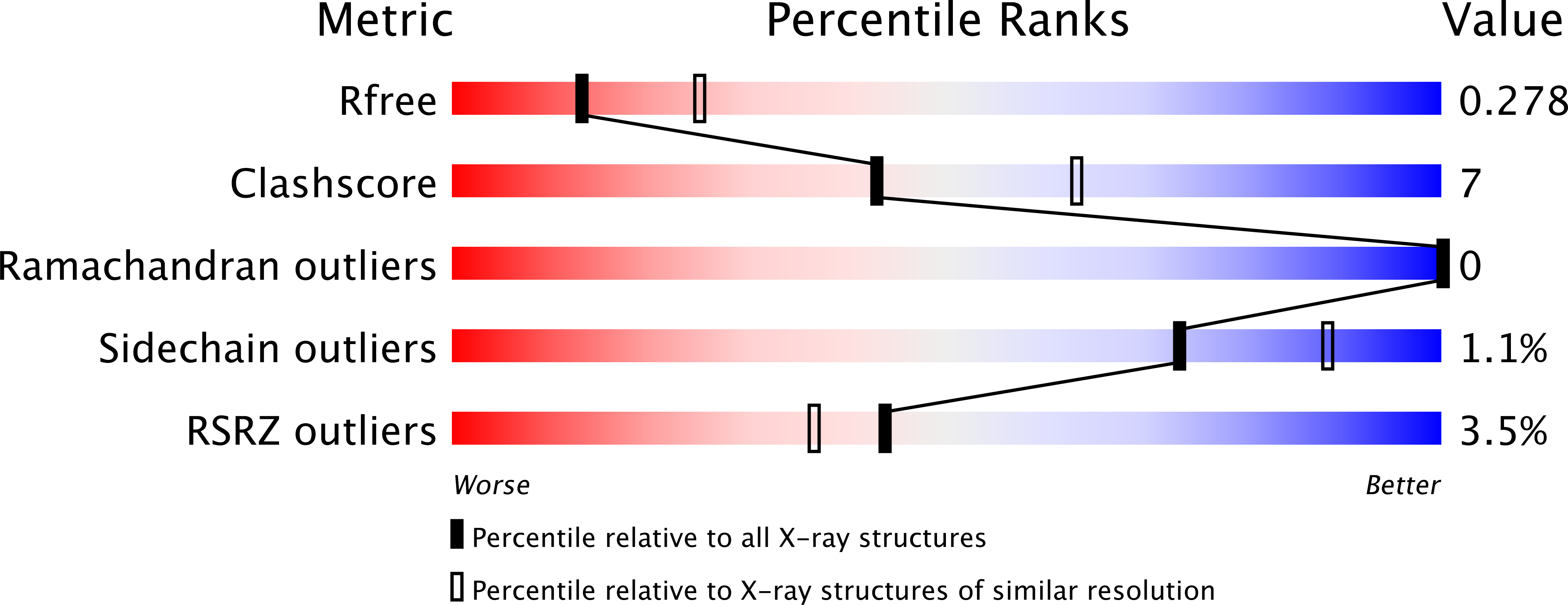
Resolution:
2.60 Å
R-Value Free:
0.27
R-Value Work:
0.25
R-Value Observed:
0.25
Space Group:
C 1 2 1
Macromolecular Entities
Structures with similar UniProt ID
Protein Blast
Polymer Type:polypeptide(L)
Molecule:Lysostaphin resistance protein A
Gene (Uniprot):lyrA
Chain IDs:A
Chain Length:278
Number of Molecules:1
Biological Source:Staphylococcus aureus (strain bovine RF122 / ET3-1)
Structures with similar UniProt ID
Protein Blast
Polymer Type:polypeptide(L)
Molecule:LYZ2 domain-containing protein
Gene (Uniprot):SAOUHSC_01895
Chain IDs:B
Chain Length:297
Number of Molecules:1
Biological Source:Staphylococcus aureus (strain NCTC 8325)
Polymer Type:polypeptide(L)
Molecule:FLAG peptide
Chain IDs:C (auth: D)
Chain Length:8
Number of Molecules:1
Biological Source:Synthetic construct
Primary Citation

Abstact

Bacteria are encapsulated by a peptidoglycan cell wall that is essential for their survival1. During cell wall assembly, a lipid-linked disaccharide-peptide precursor called lipid II is polymerized and cross-linked to produce mature peptidoglycan. As lipid II is polymerized, nascent polymers remain membrane-anchored at one end, and the other end becomes cross-linked to the matrix2-4. How bacteria release newly synthesized peptidoglycan strands from the membrane to complete the synthesis of mature peptidoglycan is a long-standing question. Here, we show that a Staphylococcus aureus cell wall hydrolase and a membrane protein that contains eight transmembrane helices form a complex that may function as a peptidoglycan release factor. The complex cleaves nascent peptidoglycan internally to produce free oligomers as well as lipid-linked oligomers that can undergo further elongation. The polytopic membrane protein, which is similar to a eukaryotic CAAX protease, controls the length of these products. A structure of the complex at a resolution of 2.6 Å shows that the membrane protein scaffolds the hydrolase to orient its active site for cleaving the glycan strand. We propose that this complex functions to detach newly synthesized peptidoglycan polymer from the cell membrane to complete integration into the cell wall matrix.

Legend

Protein

Chemical

Disease

Primary Citation of related structures
Feedback Form
Name
Email
Institute
Feedback