
Deposition Date
2019-08-09
Release Date
2020-03-11
Last Version Date
2024-11-20
Entry Detail
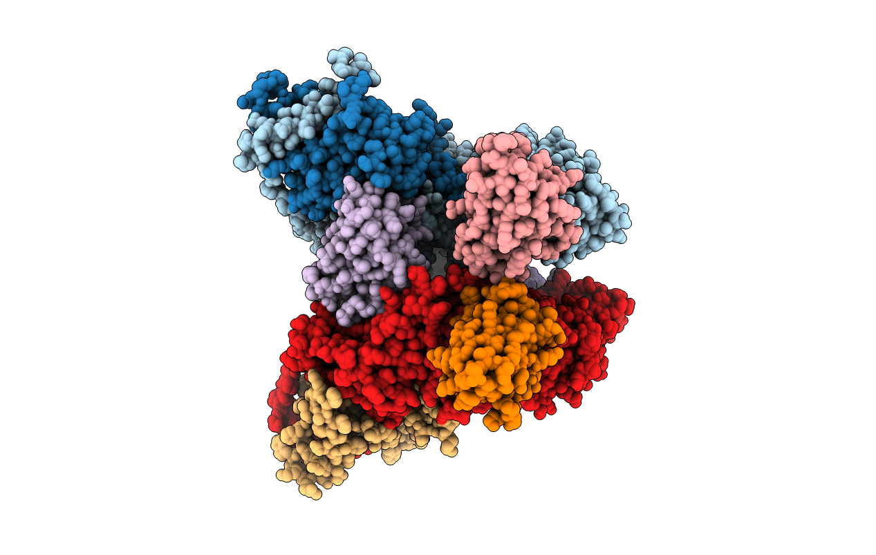
PDB ID:
6TYL
Keywords:
Title:
Crystal structure of mammalian Ric-8A:Galpha(i):nanobody complex
Biological Source:
Source Organism(s):
Rattus norvegicus (Taxon ID: 10116)
Lama glama (Taxon ID: 9844)
Lama glama (Taxon ID: 9844)
Expression System(s):
Method Details:
Experimental Method:
Resolution:
3.30 Å
R-Value Free:
0.28
R-Value Work:
0.24
Space Group:
P 1 21 1
