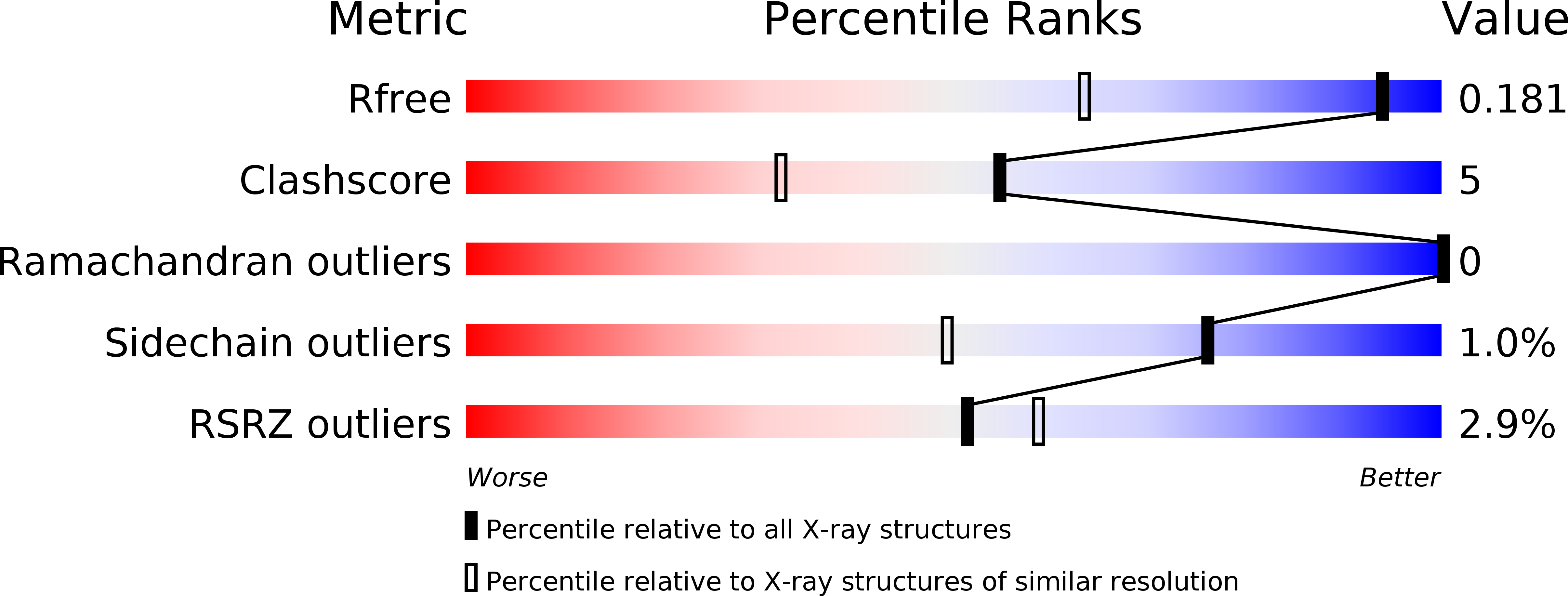
6TWO image
Deposition Date 2020-01-13
Release Date 2020-07-22
Last Version Date 2024-01-24
Entry Detail
PDB ID:
6TWO
Keywords:
Title:
Binding domain of BoNT/A6
Biological Source:
Source Organism:
Host Organism:
Method Details:
Experimental Method:
Resolution:
1.35 Å
R-Value Free:
0.17
R-Value Work:
0.14
Space Group:
P 21 21 21
Macromolecular Entities
Structures with similar UniProt ID
Protein Blast
Polymer Type:polypeptide(L)
Molecule:Bont/A1
Chain IDs:A (auth: AAA)
Chain Length:433
Number of Molecules:1
Biological Source:Clostridium botulinum
Ligand Molecules
Primary Citation
High-resolution crystal structures of the botulinum neurotoxin binding domains from subtypes A5 and A6.
Febs Open Bio 10 1474 1481 (2020)
PMID: 32654405 DOI: 10.1002/2211-5463.12931

Abstact

Clostridium botulinum neurotoxins (BoNTs) cause flaccid paralysis through inhibition of acetylcholine release from motor neurons; however, at tiny doses, this property is exploited for use as a therapeutic. Each member of the BoNT family of proteins consists of three distinct domains: a binding domain that targets neuronal cell membranes (HC), a translocation domain (HN) and a catalytic domain (LC). Here, we present high-resolution crystal structures of the binding domains of BoNT subtypes/A5 (HC /A5) and/A6 (HC /A6). These structures show that the core fold identified in other subtypes is maintained, but with subtle differences at the expected receptor-binding sites.

Legend

Protein

Chemical

Disease

Primary Citation of related structures