Deposition Date
2019-12-20
Release Date
2020-02-19
Last Version Date
2024-01-24
Entry Detail
PDB ID:
6TSJ
Keywords:
Title:
Crystal structure of human L ferritin (HuLf) Fe(III)-loaded for 15 minutes
Biological Source:
Source Organism(s):
Homo sapiens (Taxon ID: 9606)
Expression System(s):
Method Details:
Experimental Method:
Resolution:
2.30 Å
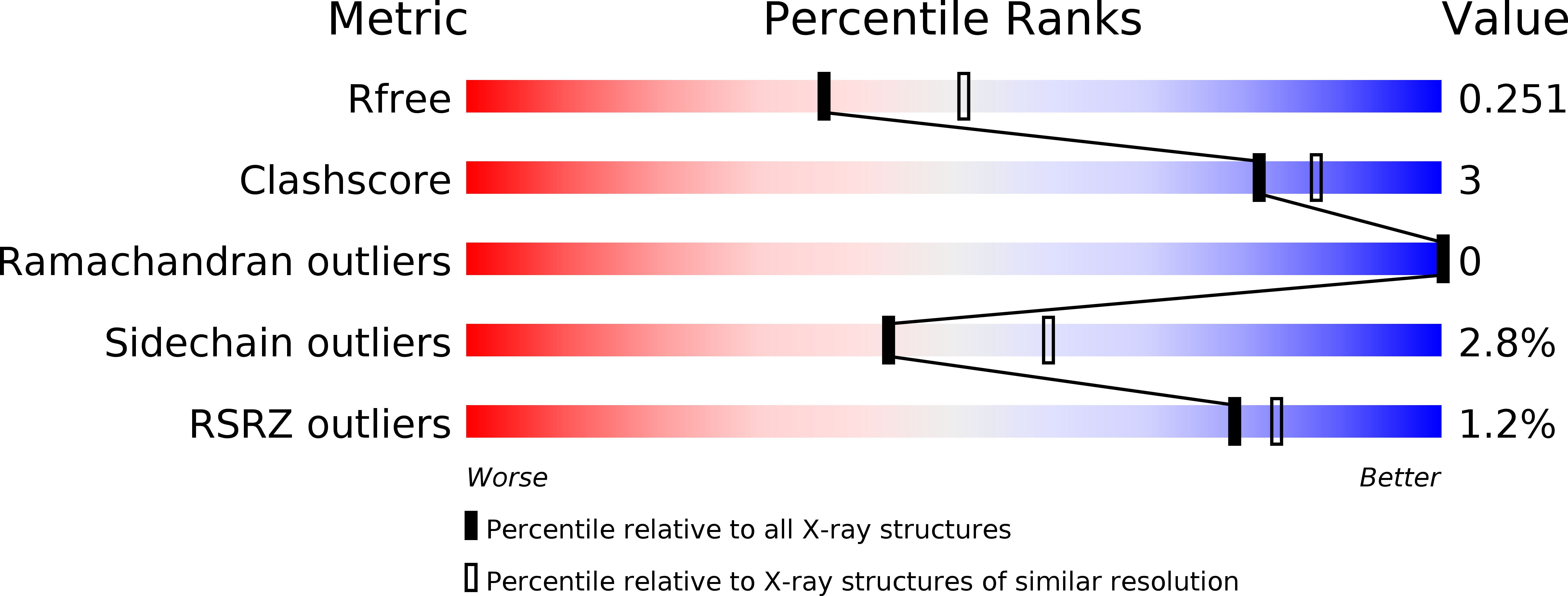
R-Value Free:
0.26
R-Value Work:
0.19
Space Group:
I 4 3 2