6TPJ image
Entry Detail
PDB ID:
6TPJ
Title:
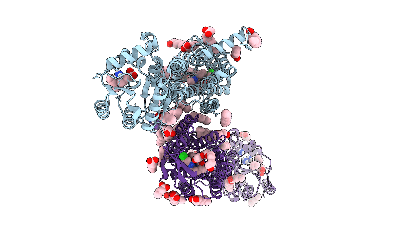
Crystal structure of the Orexin-2 receptor in complex with suvorexant at 2.76 A resolution
Biological Source:
Host Organism:
PDB Version:
Deposition Date:
2019-12-13
Release Date:
2020-01-01
Method Details:
Experimental Method:
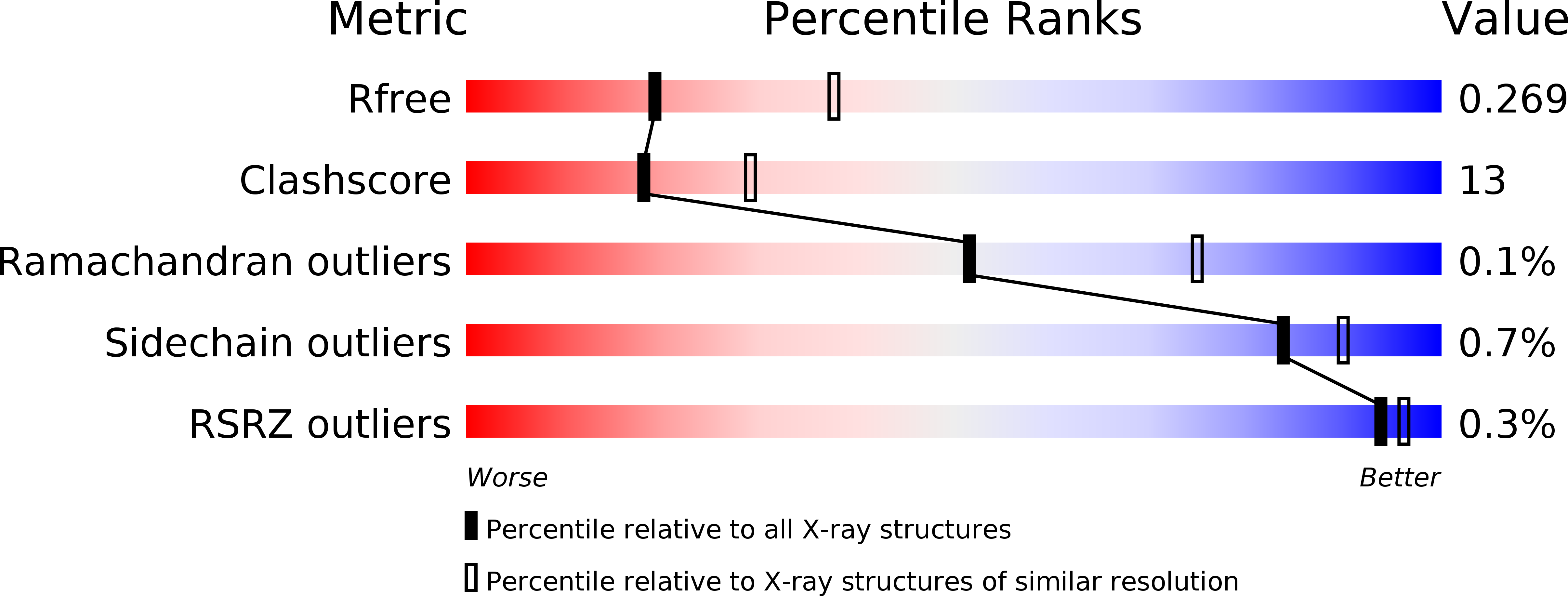
Resolution:
2.74 Å
R-Value Free:
0.25
R-Value Work:
0.20
R-Value Observed:
0.20
Space Group:
P 1
Macromolecular Entities
Polymer Type:polypeptide(L)
Description:Orexin receptor type 2,GlgA glycogen synthase,Hypocretin receptor-2
Mutations:E54A Y91L D100A V142A R170L L206A Y219A M233A A242L L310V L318A T347A N14D N22D N202D C381W C382W C383W
Chain IDs:A, B
Chain Length:564
Number of Molecules:2
Biological Source:Homo sapiens, Pyrococcus abyssi GE5
Modified Residue
Compound ID Chain ID Parent Comp ID Details 2D Image
YCM A CYS modified residue
Primary Citation
Comparison of Orexin 1 and Orexin 2 Ligand Binding Modes Using X-ray Crystallography and Computational Analysis.
J.Med.Chem. 63 1528 1543 (2020)
PMID: 31860301 DOI: 10.1021/acs.jmedchem.9b01787

Abstact

The orexin system, which consists of the two G protein-coupled receptors OX1 and OX2, activated by the neuropeptides OX-A and OX-B, is firmly established as a key regulator of behavioral arousal, sleep, and wakefulness and has been an area of intense research effort over the past two decades. X-ray structures of the receptors in complex with 10 new antagonist ligands from diverse chemotypes are presented, which complement the existing structural information for the system and highlight the critical importance of lipophilic hotspots and water molecules for these peptidergic GPCR targets. Learnings from the structural information regarding the utility of pharmacophore models and how selectivity between OX1 and OX2 can be achieved are discussed.

Legend

Protein

Chemical

Disease

Primary Citation of related structures