6TOB image
Entry Detail
PDB ID:
6TOB
Title:
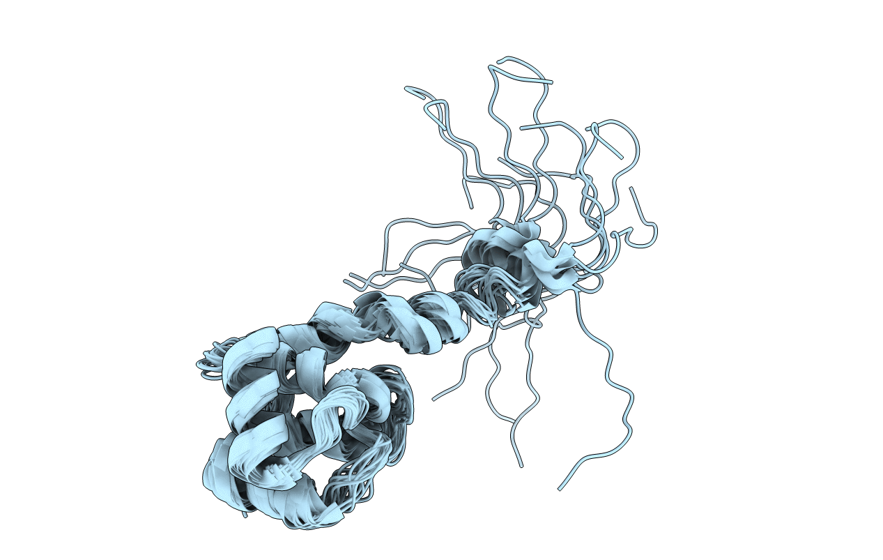
Structural and DNA Binding Properties of Mycobacterial Integration Host Factor mIHF
Biological Source:
Host Organism:
PDB Version:
Deposition Date:
2019-12-11
Release Date:
2019-12-25
Method Details:
Experimental Method:
Conformers Calculated:
80
Conformers Submitted:
20
Selection Criteria:
structures with the lowest energy
Macromolecular Entities
Polymer Type:polypeptide(L)
Description:Integration host factor MIHF
Chain IDs:A
Chain Length:109
Number of Molecules:1
Biological Source:Mycobacterium tuberculosis
Ligand Molecules
Primary Citation
Structural and DNA binding properties of mycobacterial integration host factor mIHF.
J.Struct.Biol. 209 107434 107434 (2020)
PMID: 31846718 DOI: 10.1016/j.jsb.2019.107434

Abstact

In bacteria, nucleoid associated proteins (NAPs) take part in active chromosome organization by supercoil management, three-dimensional DNA looping and direct transcriptional control. Mycobacterial integration host factor (mIHF, rv1388) is a NAP restricted to Actinobacteria and essential for survival of the human pathogen Mycobacterium tuberculosis. We show in vitro that DNA binding by mIHF strongly stabilizes the protein and increases its melting temperature. The structure obtained by Nuclear Magnetic Resonance (NMR) spectroscopy characterizes mIHF as a globular protein with a protruding alpha helix and a disordered N-terminus, similar to Streptomyces coelicolor IHF (sIHF). NMR revealed no residues of high flexibility, suggesting that mIHF is a rigid protein overall that does not undergo structural rearrangements. We show that mIHF only binds to double stranded DNA in solution, through two DNA binding sites (DBSs) similar to those identified in the X-ray structure of sIHF. According to Atomic Force Microscopy, mIHF is able to introduce left-handed loops of ca. 100 nm size (~300 bp) in supercoiled cosmids, thereby unwinding and relaxing the DNA.

Legend

Protein

Chemical

Disease

Primary Citation of related structures