6TMS image
Entry Detail
PDB ID:
6TMS
Title:
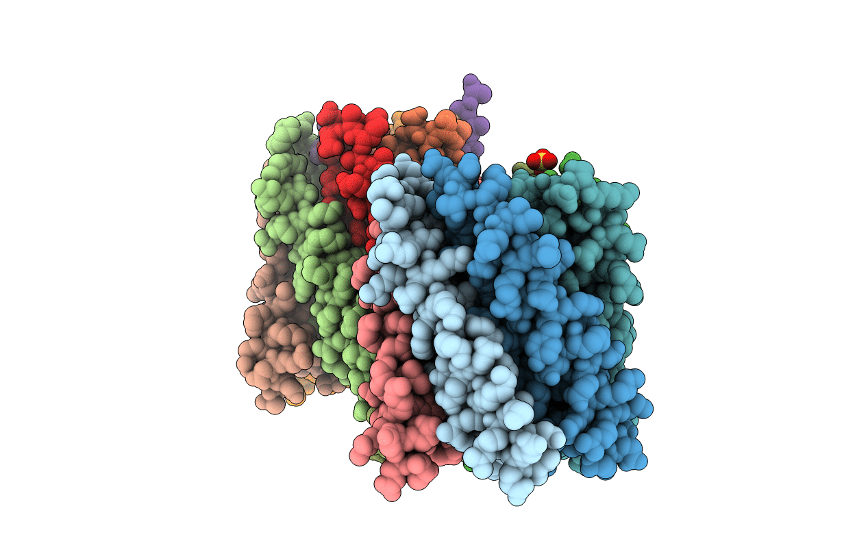
Crystal structure of a de novo designed hexameric helical-bundle protein
Biological Source:
Source Organism:
PDB Version:
Deposition Date:
2019-12-05
Release Date:
2020-04-29
Method Details:
Experimental Method:
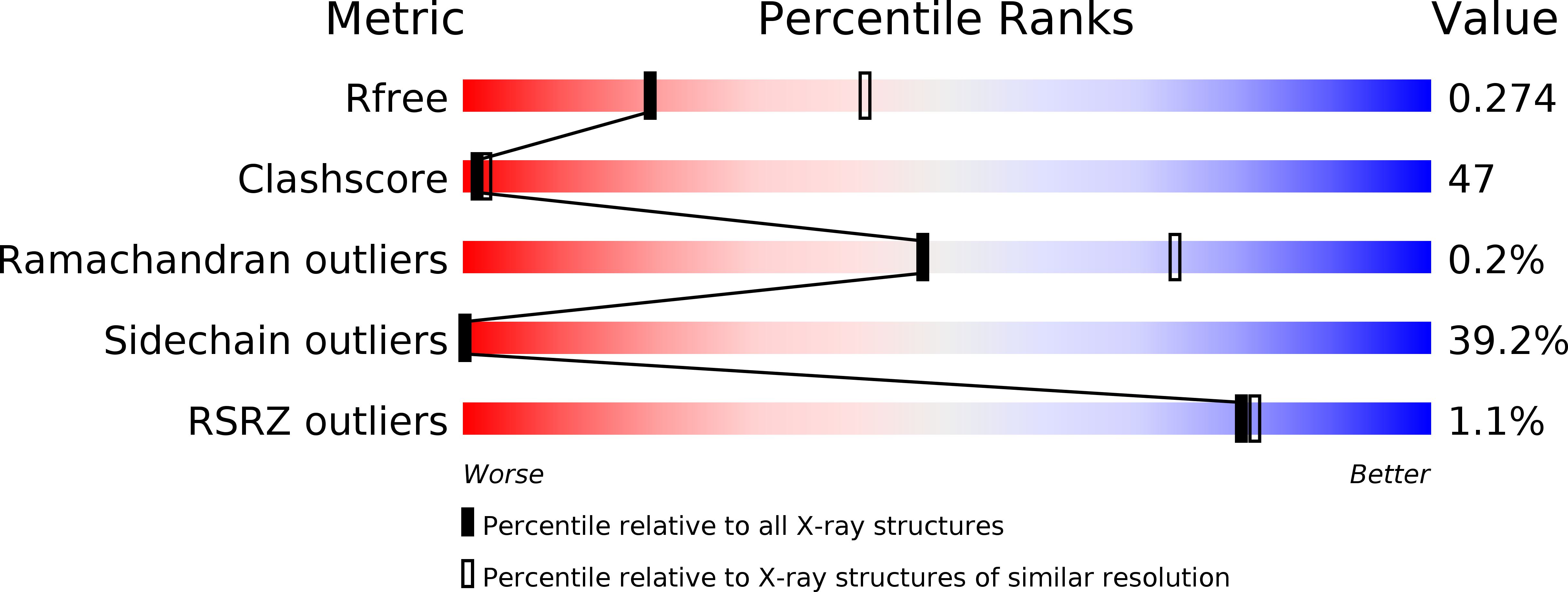
Resolution:
2.70 Å
R-Value Free:
0.29
R-Value Work:
0.28
R-Value Observed:
0.29
Space Group:
P 1
Macromolecular Entities
Polymer Type:polypeptide(L)
Description:a novel designed pore protein
Chain IDs:A, B, C, D (auth: E), E (auth: F), G (auth: D), H, I, J, K
Chain Length:69
Number of Molecules:10
Biological Source:synthetic construct
Polymer Type:polypeptide(L)
Description:a novel designed pore protein
Chain IDs:F (auth: G), L
Chain Length:69
Number of Molecules:2
Biological Source:synthetic construct
Polymer Type:polypeptide(L)
Description:affinity purification tag
Chain IDs:M (auth: Q), N (auth: R)
Chain Length:16
Number of Molecules:2
Biological Source:synthetic construct
Ligand Molecules
Primary Citation

Abstact

Transmembrane channels and pores have key roles in fundamental biological processes1 and in biotechnological applications such as DNA nanopore sequencing2-4, resulting in considerable interest in the design of pore-containing proteins. Synthetic amphiphilic peptides have been found to form ion channels5,6, and there have been recent advances in de novo membrane protein design7,8 and in redesigning naturally occurring channel-containing proteins9,10. However, the de novo design of stable, well-defined transmembrane protein pores that are capable of conducting ions selectively or are large enough to enable the passage of small-molecule fluorophores remains an outstanding challenge11,12. Here we report the computational design of protein pores formed by two concentric rings of α-helices that are stable and monodisperse in both their water-soluble and their transmembrane forms. Crystal structures of the water-soluble forms of a 12-helical pore and a 16-helical pore closely match the computational design models. Patch-clamp electrophysiology experiments show that, when expressed in insect cells, the transmembrane form of the 12-helix pore enables the passage of ions across the membrane with high selectivity for potassium over sodium; ion passage is blocked by specific chemical modification at the pore entrance. When incorporated into liposomes using in vitro protein synthesis, the transmembrane form of the 16-helix pore-but not the 12-helix pore-enables the passage of biotinylated Alexa Fluor 488. A cryo-electron microscopy structure of the 16-helix transmembrane pore closely matches the design model. The ability to produce structurally and functionally well-defined transmembrane pores opens the door to the creation of designer channels and pores for a wide variety of applications.

Legend

Protein

Chemical

Disease

Primary Citation of related structures