6THY image
Deposition Date 2019-11-21
Release Date 2020-01-29
Last Version Date 2024-01-24
Entry Detail
PDB ID:
6THY
Keywords:
Title:
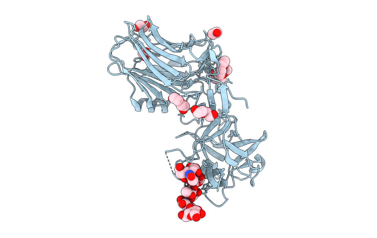
Botulinum neurotoxin A3 Hc domain in complex with GD1a
Biological Source:
Source Organism:
Host Organism:
Method Details:
Experimental Method:
Resolution:
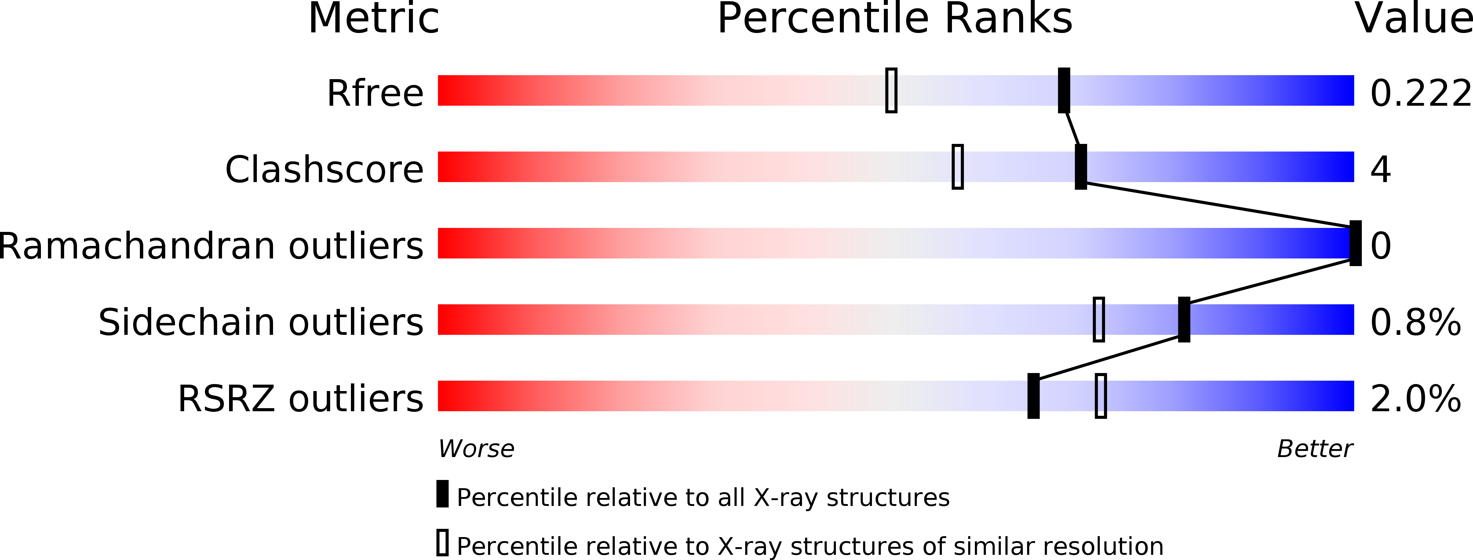
1.75 Å
R-Value Free:
0.21
R-Value Work:
0.17
Space Group:
P 21 21 21
Macromolecular Entities
Structures with similar UniProt ID
Protein Blast
Polymer Type:polypeptide(L)
Molecule:BoNT/A3
Chain IDs:A (auth: AAA)
Chain Length:433
Number of Molecules:1
Biological Source:Clostridium botulinum
Primary Citation
Crystal structure of botulinum neurotoxin subtype A3 cell binding domain in complex with GD1a co-receptor ganglioside.
Febs Open Bio 10 298 305 (2020)
PMID: 31945264 DOI: 10.1002/2211-5463.12790

Abstact

Botulinum neurotoxins (BoNTs) are one of the most toxic proteins known to humans. Their molecular structure is comprised of three essential domains-a cell binding domain (HC), translocation domain and catalytic domain (light chain) . The HC domain facilitates the highly specific binding of BoNTs to the neuronal membrane via a dual-receptor complex involving a protein receptor and a ganglioside. Variation in activity/toxicity across subtypes of serotype A has been attributed to changes in protein and ganglioside interactions, and their implications are important in the design of novel BoNT-based therapeutics. Here, we present the structure of BoNT/A3 cell binding domain (HC /A3) in complex with the ganglioside GD1a at 1.75 Å resolution. The structure revealed that six residues interact with the three outermost monosaccharides of GD1a through several key hydrogen bonding interactions. A detailed comparison of structures of HC /A3 with HC /A1 revealed subtle conformational differences at the ganglioside binding site upon carbohydrate binding.

Legend

Protein

Chemical

Disease

Primary Citation of related structures