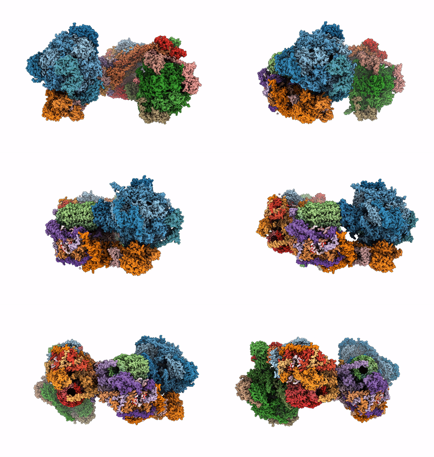
6TDU image
Entry Detail
PDB ID:
6TDU
EMDB ID:
Title:
Cryo-EM structure of Euglena gracilis mitochondrial ATP synthase, full dimer, rotational states 1
Biological Source:
Source Organism:
PDB Version:
Deposition Date:
2019-11-10
Release Date:
2019-11-27
Method Details:
Experimental Method:
Resolution:
4.32 Å
Aggregation State:
PARTICLE
Reconstruction Method:
SINGLE PARTICLE
Macromolecular Entities
Polymer Type:polypeptide(L)
Description:ATPTB1
Chain IDs:A, S (auth: a)
Chain Length:487
Number of Molecules:2
Biological Source:Euglena gracilis
Polymer Type:polypeptide(L)
Description:ATP synthase subunit alpha
Chain IDs:KA (auth: AA), LA (auth: AB), MA (auth: AC), KB (auth: BA), LB (auth: BB), MB (auth: BC)
Chain Length:561
Number of Molecules:6
Biological Source:Euglena gracilis
Polymer Type:polypeptide(L)
Description:ATP synthase subunit beta
Chain IDs:NA (auth: AD), OA (auth: AE), PA (auth: AF), NB (auth: BD), OB (auth: BE), PB (auth: BF)
Chain Length:501
Number of Molecules:6
Biological Source:Euglena gracilis
Polymer Type:polypeptide(L)
Description:ATP synthase subunit gamma
Chain IDs:QA (auth: AG), QB (auth: BG)
Chain Length:306
Number of Molecules:2
Biological Source:Euglena gracilis
Polymer Type:polypeptide(L)
Description:ATP synthase subunit delta
Chain IDs:RA (auth: AH), RB (auth: BH)
Chain Length:176
Number of Molecules:2
Biological Source:Euglena gracilis
Polymer Type:polypeptide(L)
Description:ATP synthase subunit epsilon
Chain IDs:SA (auth: AI), SB (auth: BI)
Chain Length:76
Number of Molecules:2
Biological Source:Euglena gracilis
Polymer Type:polypeptide(L)
Description:p18
Chain IDs:TA (auth: AJ), UA (auth: AK), VA (auth: AL), TB (auth: BJ), UB (auth: BK), VB (auth: BL)
Chain Length:192
Number of Molecules:6
Biological Source:Euglena gracilis
Polymer Type:polypeptide(L)
Description:oligomycin sensitivity conferring protein (OSCP)
Chain IDs:WA (auth: AM), WB (auth: BM)
Chain Length:267
Number of Molecules:2
Biological Source:Euglena gracilis
Polymer Type:polypeptide(L)
Description:inhibitor of F1 (IF1)
Chain IDs:XA (auth: AN), XB (auth: BN)
Chain Length:103
Number of Molecules:2
Biological Source:Euglena gracilis
Polymer Type:polypeptide(L)
Description:ATP synthase subunit c
Chain IDs:YA (auth: AO), ZA (auth: AP), AB (auth: AQ), BB (auth: AR), CB (auth: AS), DB (auth: AT), EB (auth: AU), FB (auth: AV), GB (auth: AW), HB (auth: AX), YB (auth: BO), ZB (auth: BP), AC (auth: BQ), BC (auth: BR), CC (auth: BS), DC (auth: BT), EC (auth: BU), FC (auth: BV), GC (auth: BW), HC (auth: BX)
Chain Length:104
Number of Molecules:20
Biological Source:Euglena gracilis
Polymer Type:polypeptide(L)
Description:ATPTB3
Chain IDs:IB (auth: B), IC (auth: b)
Chain Length:338
Number of Molecules:2
Biological Source:Euglena gracilis
Polymer Type:polypeptide(L)
Description:ATPTB4
Chain IDs:JB (auth: C), JC (auth: c)
Chain Length:169
Number of Molecules:2
Biological Source:Euglena gracilis
Polymer Type:polypeptide(L)
Description:ATPTB6
Chain IDs:B (auth: D), T (auth: d)
Chain Length:187
Number of Molecules:2
Biological Source:Euglena gracilis
Polymer Type:polypeptide(L)
Description:ATPTB12
Chain IDs:C (auth: E), U (auth: e)
Chain Length:97
Number of Molecules:2
Biological Source:Euglena gracilis
Polymer Type:polypeptide(L)
Description:ATP synthase subunit a
Chain IDs:D (auth: F), V (auth: f)
Chain Length:274
Number of Molecules:2
Biological Source:Euglena gracilis
Polymer Type:polypeptide(L)
Description:ATP synthase subunit b
Chain IDs:E (auth: G), W (auth: g)
Chain Length:112
Number of Molecules:2
Biological Source:Euglena gracilis
Polymer Type:polypeptide(L)
Description:ATP synthase subunit d
Chain IDs:F (auth: H), X (auth: h)
Chain Length:476
Number of Molecules:2
Biological Source:Euglena gracilis
Polymer Type:polypeptide(L)
Description:ATP synthase subunit f
Chain IDs:G (auth: I), Y (auth: i)
Chain Length:98
Number of Molecules:2
Biological Source:Euglena gracilis
Polymer Type:polypeptide(L)
Description:ATP synthase subunit i/j
Chain IDs:H (auth: J), Z (auth: j)
Chain Length:104
Number of Molecules:2
Biological Source:Euglena gracilis
Polymer Type:polypeptide(L)
Description:ATP synthase subunit k
Chain IDs:I (auth: K), AA (auth: k)
Chain Length:113
Number of Molecules:2
Biological Source:Euglena gracilis
Polymer Type:polypeptide(L)
Description:ATP synthase subunit 8
Chain IDs:J (auth: L), BA (auth: l)
Chain Length:57
Number of Molecules:2
Biological Source:Euglena gracilis
Polymer Type:polypeptide(L)
Description:ATPEG1
Chain IDs:K (auth: M), CA (auth: m)
Chain Length:169
Number of Molecules:2
Biological Source:Euglena gracilis
Polymer Type:polypeptide(L)
Description:ATPEG2
Chain IDs:L (auth: N), DA (auth: n)
Chain Length:137
Number of Molecules:2
Biological Source:Euglena gracilis
Polymer Type:polypeptide(L)
Description:ATPEG3
Chain IDs:M (auth: O), EA (auth: o)
Chain Length:116
Number of Molecules:2
Biological Source:Euglena gracilis
Polymer Type:polypeptide(L)
Description:ATPEG4
Chain IDs:N (auth: P), FA (auth: p)
Chain Length:120
Number of Molecules:2
Biological Source:Euglena gracilis
Polymer Type:polypeptide(L)
Description:ATPEG5
Chain IDs:O (auth: Q), GA (auth: q)
Chain Length:90
Number of Molecules:2
Biological Source:Euglena gracilis
Polymer Type:polypeptide(L)
Description:ATPEG6
Chain IDs:P (auth: R), HA (auth: r)
Chain Length:78
Number of Molecules:2
Biological Source:Euglena gracilis
Polymer Type:polypeptide(L)
Description:ATPEG7
Chain IDs:Q (auth: S), IA (auth: s)
Chain Length:74
Number of Molecules:2
Biological Source:Euglena gracilis
Polymer Type:polypeptide(L)
Description:ATPEG8
Chain IDs:R (auth: T), JA (auth: t)
Chain Length:66
Number of Molecules:2
Biological Source:Euglena gracilis
Primary Citation
Structure of a mitochondrial ATP synthase with bound native cardiolipin.
Elife 8 ? ? (2019)
PMID: 31738165 DOI: 10.7554/eLife.51179

Abstact

The mitochondrial ATP synthase fuels eukaryotic cells with chemical energy. Here we report the cryo-EM structure of a divergent ATP synthase dimer from mitochondria of Euglena gracilis, a member of the phylum Euglenozoa that also includes human parasites. It features 29 different subunits, 8 of which are newly identified. The membrane region was determined to 2.8 Å resolution, enabling the identification of 37 associated lipids, including 25 cardiolipins, which provides insight into protein-lipid interactions and their functional roles. The rotor-stator interface comprises four membrane-embedded horizontal helices, including a distinct subunit a. The dimer interface is formed entirely by phylum-specific components, and a peripherally associated subcomplex contributes to the membrane curvature. The central and peripheral stalks directly interact with each other. Last, the ATPase inhibitory factor 1 (IF1) binds in a mode that is different from human, but conserved in Trypanosomatids.

Legend

Protein

Chemical

Disease

Primary Citation of related structures