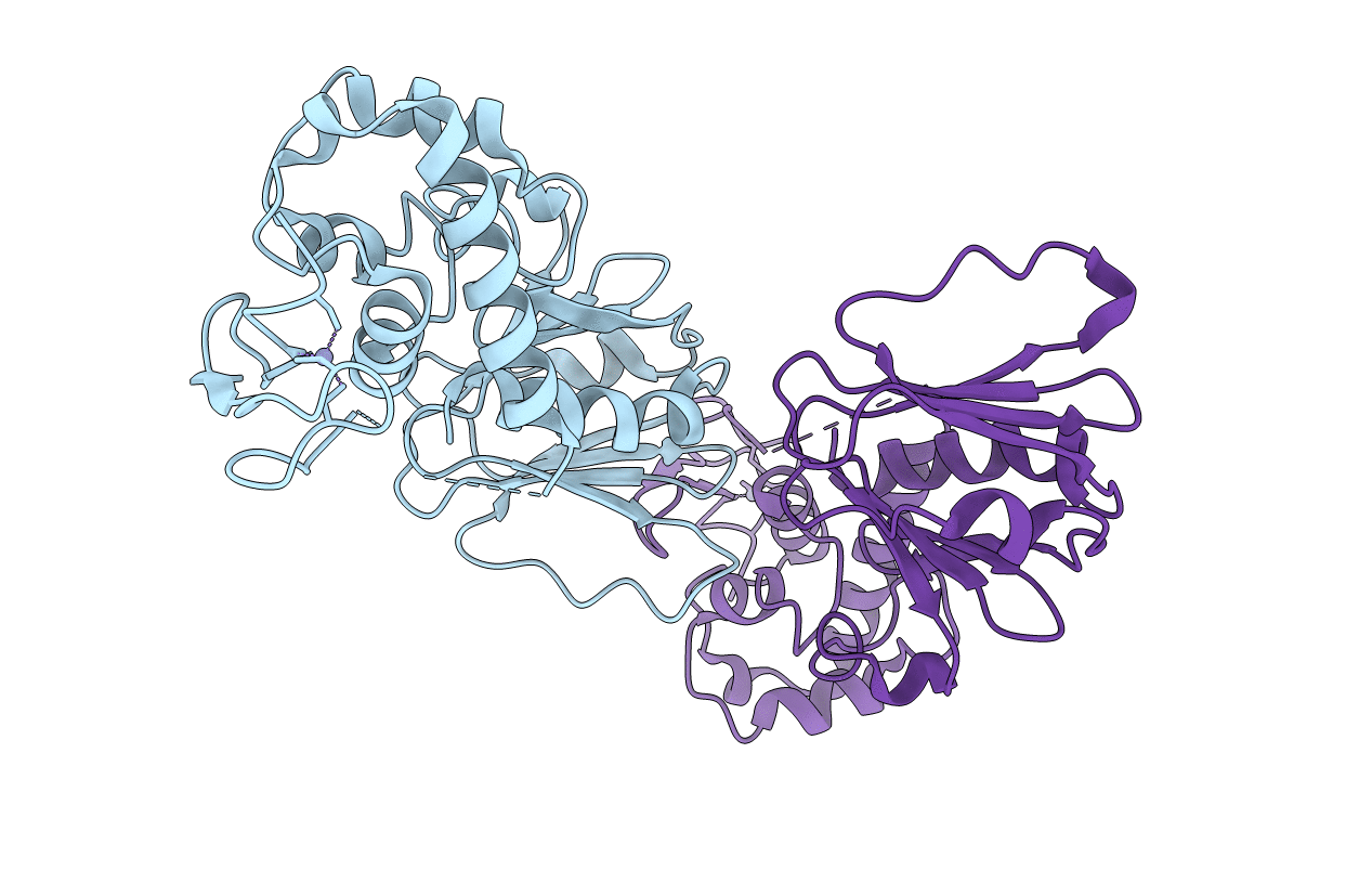
6TC6 image
Deposition Date 2019-11-05
Release Date 2020-10-14
Last Version Date 2024-05-15
Entry Detail
PDB ID:
6TC6
Keywords:
Title:
Crystal structure of MutM from Neisseria meningitidis
Biological Source:
Source Organism:
Host Organism:
Method Details:
Experimental Method:
Resolution:
2.90 Å
R-Value Free:
0.29
R-Value Work:
0.24
R-Value Observed:
0.24
Space Group:
P 1 2 1
Macromolecular Entities
Polymer Type:polypeptide(L)
Molecule:Formamidopyrimidine-DNA glycosylase
Gene (Uniprot):mutM
Chain IDs:A, B (auth: C)
Chain Length:275
Number of Molecules:2
Biological Source:Neisseria meningitidis alpha522
Ligand Molecules
Primary Citation
Conformational changes of DNA repair glycosylase MutM triggered by DNA binding.
Febs Lett. 594 3032 3044 (2020)
PMID: 32598485 DOI: 10.1002/1873-3468.13876

Abstact

Bacterial MutM is a DNA repair glycosylase removing DNA damage generated from oxidative stress and, therefore, preventing mutations and genomic instability. MutM belongs to the Fpg/Nei family of prokaryotic enzymes sharing structural and functional similarities with their eukaryotic counterparts, for example, NEIL1-NEIL3. Here, we present two crystal structures of MutM from pathogenic Neisseria meningitidis: a MutM holoenzyme and MutM bound to DNA. The free enzyme exists in an open conformation, while upon binding to DNA, both the enzyme and DNA undergo substantial structural changes and domain rearrangement. Our data show that not only NEI glycosylases but also the MutMs undergo dramatic conformational changes. Moreover, crystallographic data support the previously published observations that MutM enzymes are rather flexible and dynamic molecules.

Legend

Protein

Chemical

Disease

Primary Citation of related structures