6T7E image
Deposition Date 2019-10-21
Release Date 2020-07-22
Last Version Date 2024-01-24
Entry Detail
PDB ID:
6T7E
Title:
PII-like protein CutA from Nostoc sp. PCC7120 in complex with MES
Biological Source:
Method Details:
Experimental Method:
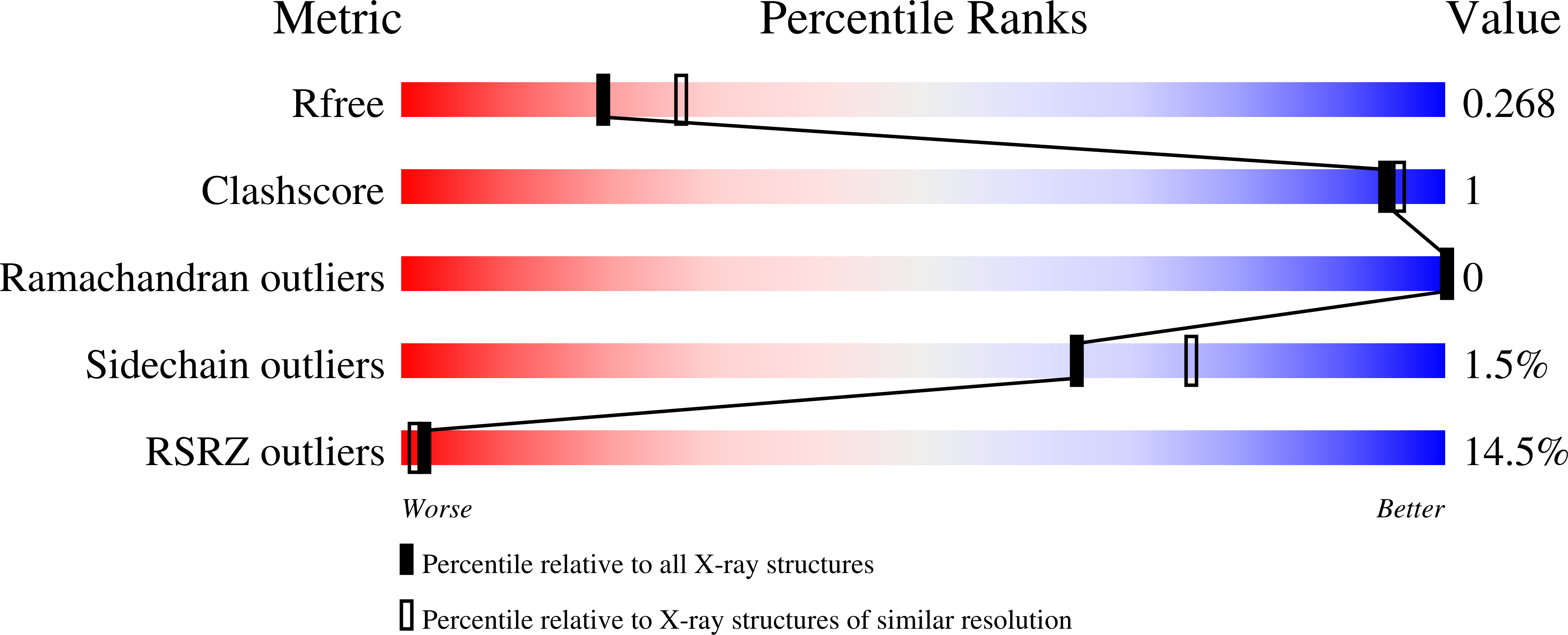
Resolution:
2.45 Å
R-Value Free:
0.27
R-Value Work:
0.22
Space Group:
P 3 2 1
Macromolecular Entities
Polymer Type:polypeptide(L)
Molecule:Periplasmic divalent cation tolerance protein
Gene (Uniprot):alr7093
Chain IDs:A, B, C, D, E, F
Chain Length:114
Number of Molecules:6
Biological Source:Nostoc sp. (strain PCC 7120 / SAG 25.82 / UTEX 2576)
Ligand Molecules
Primary Citation
Functional and structural characterization of PII-like protein CutA does not support involvement in heavy metal tolerance and hints at a small-molecule carrying/signaling role.
Febs J. 288 1142 1162 (2021)
PMID: 32599651 DOI: 10.1111/febs.15464

Abstact

The PII-like protein CutA is annotated as being involved in Cu2+ tolerance, based on analysis of Escherichia coli mutants. However, the precise cellular function of CutA remains unclear. Our bioinformatic analysis reveals that CutA proteins are universally distributed across all domains of life. Based on sequence-based clustering, we chose representative cyanobacterial CutA proteins for physiological, biochemical, and structural characterization and examined their involvement in heavy metal tolerance, by generating CutA mutants in filamentous Nostoc sp. and in unicellular Synechococcus elongatus. However, we were unable to find any involvement of cyanobacterial CutA in metal tolerance under various conditions. This prompted us to re-examine experimentally the role of CutA in protecting E. coli from Cu2+ . Since we found no effect on copper tolerance, we conclude that CutA plays a different role that is not involved in metal protection. We resolved high-resolution CutA structures from Nostoc and S. elongatus. Similarly to their counterpart from E. coli and to canonical PII proteins, cyanobacterial CutA proteins are trimeric in solution and in crystal structure; however, no binding affinity for small signaling molecules or for Cu2+ could be detected. The clefts between the CutA subunits, corresponding to the binding pockets of PII proteins, are formed by conserved aromatic and charged residues, suggesting a conserved binding/signaling function for CutA. In fact, we find binding of organic Bis-Tris/MES molecules in CutA crystal structures, revealing a strong tendency of these pockets to accommodate cargo. This highlights the need to search for the potential physiological ligands and for their signaling functions upon binding to CutA. DATABASES: Structural data are available in Protein Data Bank (PDB) under the accession numbers 6GDU, 6GDV, 6GDW, 6GDX, 6T76, and 6T7E.

Legend

Protein

Chemical

Disease

Primary Citation of related structures