
Deposition Date
2019-10-21
Release Date
2020-01-15
Last Version Date
2024-05-22
Entry Detail
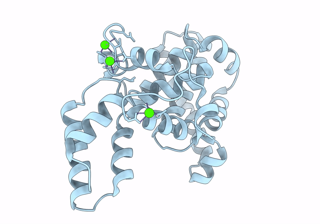
PDB ID:
6T72
Keywords:
Title:
Structure of the RsaA N-terminal domain bound to LPS
Biological Source:
Source Organism(s):
Caulobacter vibrioides CB15 (Taxon ID: 190650)
Expression System(s):
Method Details:
Experimental Method:
Resolution:
3.70 Å
Aggregation State:
PARTICLE
Reconstruction Method:
SINGLE PARTICLE
