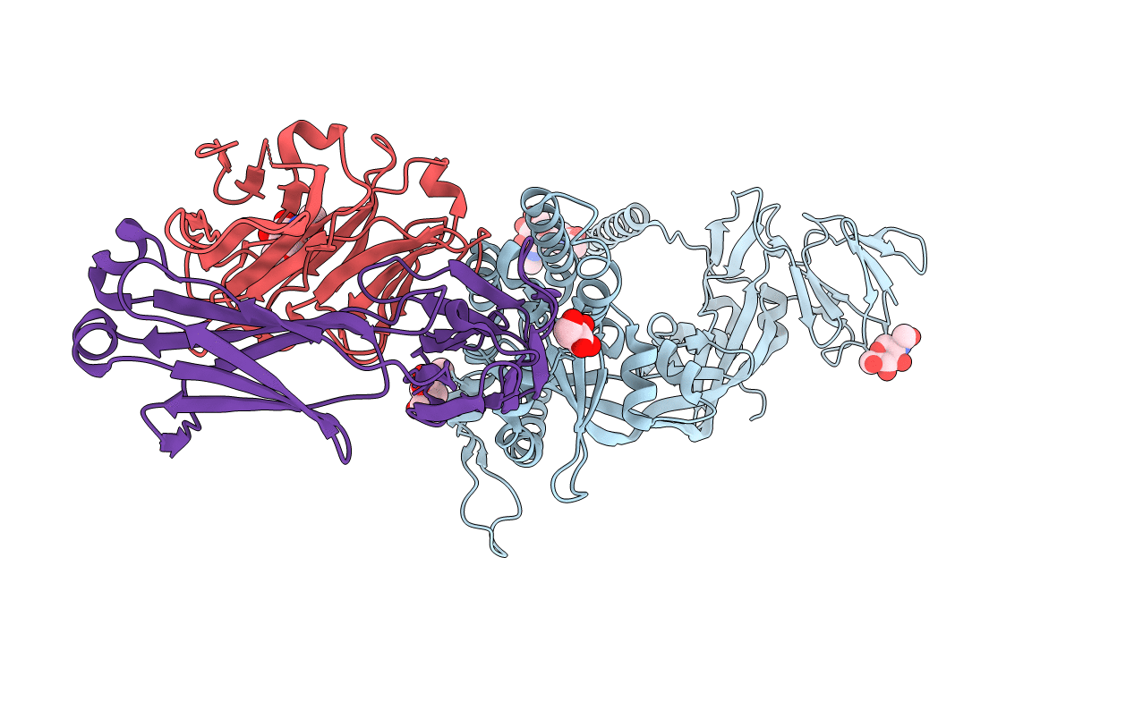
6T3F image
Deposition Date 2019-10-10
Release Date 2019-11-27
Last Version Date 2024-11-20
Entry Detail
PDB ID:
6T3F
Keywords:
Title:
Crystal structure Nipah virus fusion glycoprotein in complex with a neutralising Fab fragment
Biological Source:
Source Organism:
Host Organism:
Method Details:
Experimental Method:
Resolution:
3.20 Å
R-Value Free:
0.24
R-Value Work:
0.21
R-Value Observed:
0.21
Space Group:
P 63 2 2
Macromolecular Entities
Structures with similar UniProt ID
Protein Blast
Polymer Type:polypeptide(L)
Molecule:Fusion glycoprotein F0
Gene (Uniprot):F
Chain IDs:A (auth: F)
Chain Length:502
Number of Molecules:1
Biological Source:Nipah virus
Polymer Type:polypeptide(L)
Molecule:Fab66 heavy chain
Chain IDs:C (auth: H)
Chain Length:228
Number of Molecules:1
Biological Source:Oryctolagus cuniculus
Polymer Type:polypeptide(L)
Molecule:Fab66 light chain
Chain IDs:B (auth: L)
Chain Length:216
Number of Molecules:1
Biological Source:Oryctolagus cuniculus
Ligand Molecules
Primary Citation
A structural basis for antibody-mediated neutralization of Nipah virus reveals a site of vulnerability at the fusion glycoprotein apex.
Proc.Natl.Acad.Sci.USA 116 25057 25067 (2019)
PMID: 31767754 DOI: 10.1073/pnas.1912503116

Abstact

Nipah virus (NiV) is a highly pathogenic paramyxovirus that causes frequent outbreaks of severe neurologic and respiratory disease in humans with high case fatality rates. The 2 glycoproteins displayed on the surface of the virus, NiV-G and NiV-F, mediate host-cell attachment and membrane fusion, respectively, and are targets of the host antibody response. Here, we provide a molecular basis for neutralization of NiV through antibody-mediated targeting of NiV-F. Structural characterization of a neutralizing antibody (nAb) in complex with trimeric prefusion NiV-F reveals an epitope at the membrane-distal domain III (DIII) of the molecule, a region that undergoes substantial refolding during host-cell entry. The epitope of this monoclonal antibody (mAb66) is primarily protein-specific and we observe that glycosylation at the periphery of the interface likely does not inhibit mAb66 binding to NiV-F. Further characterization reveals that a Hendra virus-F-specific nAb (mAb36) and many antibodies in an antihenipavirus-F polyclonal antibody mixture (pAb835) also target this region of the molecule. Integrated with previously reported paramyxovirus F-nAb structures, these data support a model whereby the membrane-distal region of the F protein is targeted by the antibody-mediated immune response across henipaviruses. Notably, our domain-specific sequence analysis reveals no evidence of selective pressure at this region of the molecule, suggestive that functional constraints prevent immune-driven sequence variation. Combined, our data reveal the membrane-distal region of NiV-F as a site of vulnerability on the NiV surface.

Legend

Protein

Chemical

Disease

Primary Citation of related structures