Deposition Date
2019-09-27
Release Date
2020-05-13
Last Version Date
2024-11-13
Entry Detail
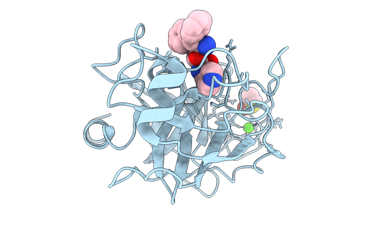
PDB ID:
6SY3
Keywords:
Title:
Cationic Trypsin in Complex with a D-DiPhe-Pro-pyridine derivative
Biological Source:
Source Organism(s):
Bos taurus (Taxon ID: 9913)
Method Details:
Experimental Method:
Resolution:
0.95 Å
R-Value Free:
0.13
R-Value Work:
0.12
R-Value Observed:
0.12
Space Group:
P 21 21 21