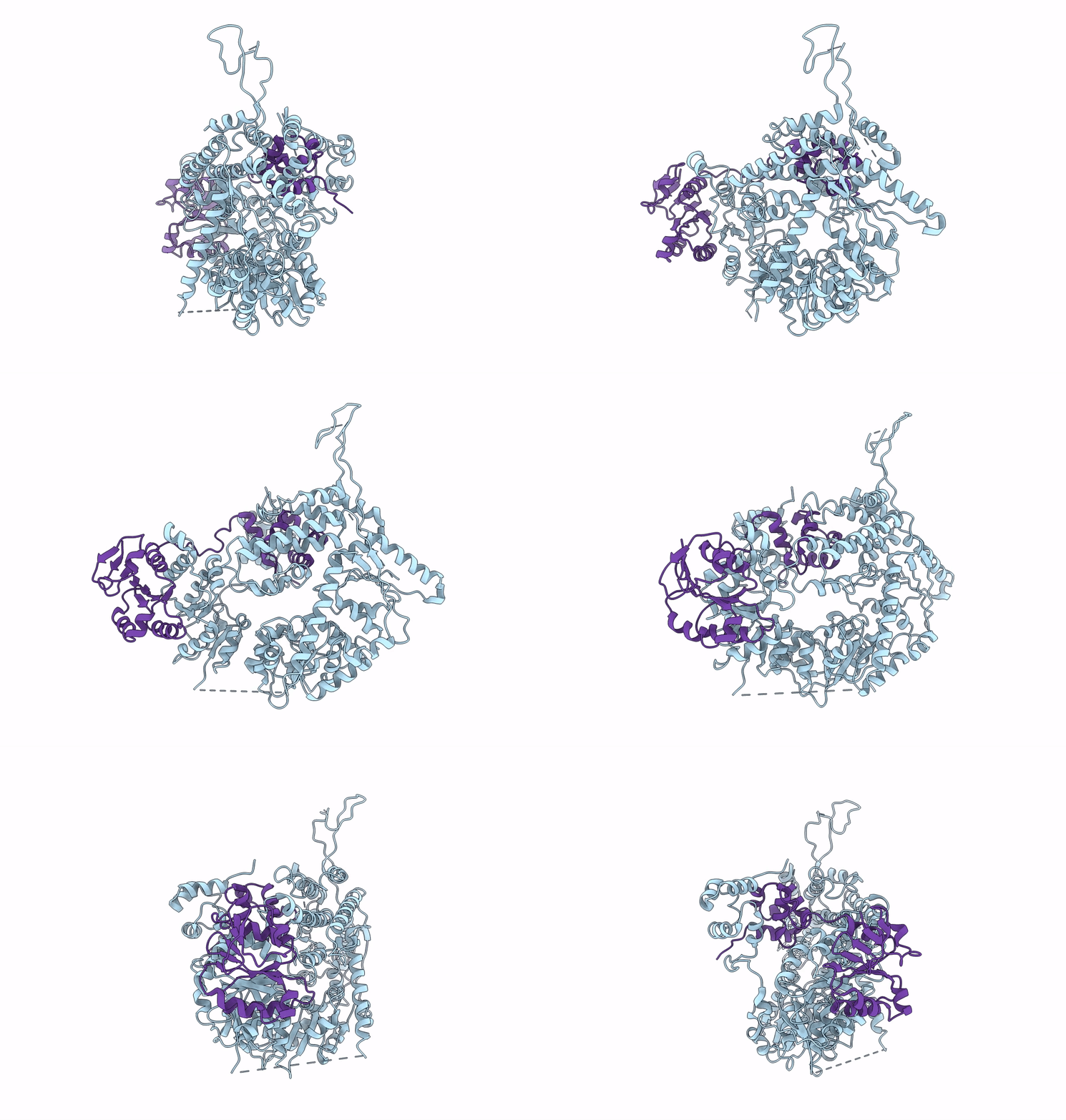
6SXA image
Entry Detail
PDB ID:
6SXA
EMDB ID:
Title:
XPF-ERCC1 Cryo-EM Structure, Apo-form
Biological Source:
Source Organism:
Host Organism:
PDB Version:
Deposition Date:
2019-09-25
Release Date:
2020-03-11
Method Details:
Experimental Method:
Resolution:
3.60 Å
Aggregation State:
2D ARRAY
Reconstruction Method:
SINGLE PARTICLE
Macromolecular Entities
Polymer Type:polypeptide(L)
Description:DNA repair endonuclease XPF
Chain IDs:A (auth: F)
Chain Length:916
Number of Molecules:1
Biological Source:Homo sapiens
Polymer Type:polypeptide(L)
Description:DNA excision repair protein ERCC-1
Chain IDs:B (auth: G)
Chain Length:297
Number of Molecules:1
Biological Source:Homo sapiens
Ligand Molecules
Primary Citation
Cryo-EM structures of the XPF-ERCC1 endonuclease reveal how DNA-junction engagement disrupts an auto-inhibited conformation.
Nat Commun 11 1120 1120 (2020)
PMID: 32111838 DOI: 10.1038/s41467-020-14856-2

Abstact

The structure-specific endonuclease XPF-ERCC1 participates in multiple DNA damage repair pathways including nucleotide excision repair (NER) and inter-strand crosslink repair (ICLR). How XPF-ERCC1 is catalytically activated by DNA junction substrates is not currently understood. Here we report cryo-electron microscopy structures of both DNA-free and DNA-bound human XPF-ERCC1. DNA-free XPF-ERCC1 adopts an auto-inhibited conformation in which the XPF helical domain masks the ERCC1 (HhH)2 domain and restricts access to the XPF catalytic site. DNA junction engagement releases the ERCC1 (HhH)2 domain to couple with the XPF-ERCC1 nuclease/nuclease-like domains. Structure-function data indicate xeroderma pigmentosum patient mutations frequently compromise the structural integrity of XPF-ERCC1. Fanconi anaemia patient mutations in XPF often display substantial in-vitro activity but are resistant to activation by ICLR recruitment factor SLX4. Our data provide insights into XPF-ERCC1 architecture and catalytic activation.

Legend

Protein

Chemical

Disease

Primary Citation of related structures