Deposition Date
2019-09-20
Release Date
2020-08-05
Last Version Date
2024-05-15
Entry Detail
PDB ID:
6SWG
Keywords:
Title:
Crystal structure of the TASOR-Periphilin core complex
Biological Source:
Source Organism(s):
Homo sapiens (Taxon ID: 9606)
Expression System(s):
Method Details:
Experimental Method:
Resolution:
2.51 Å
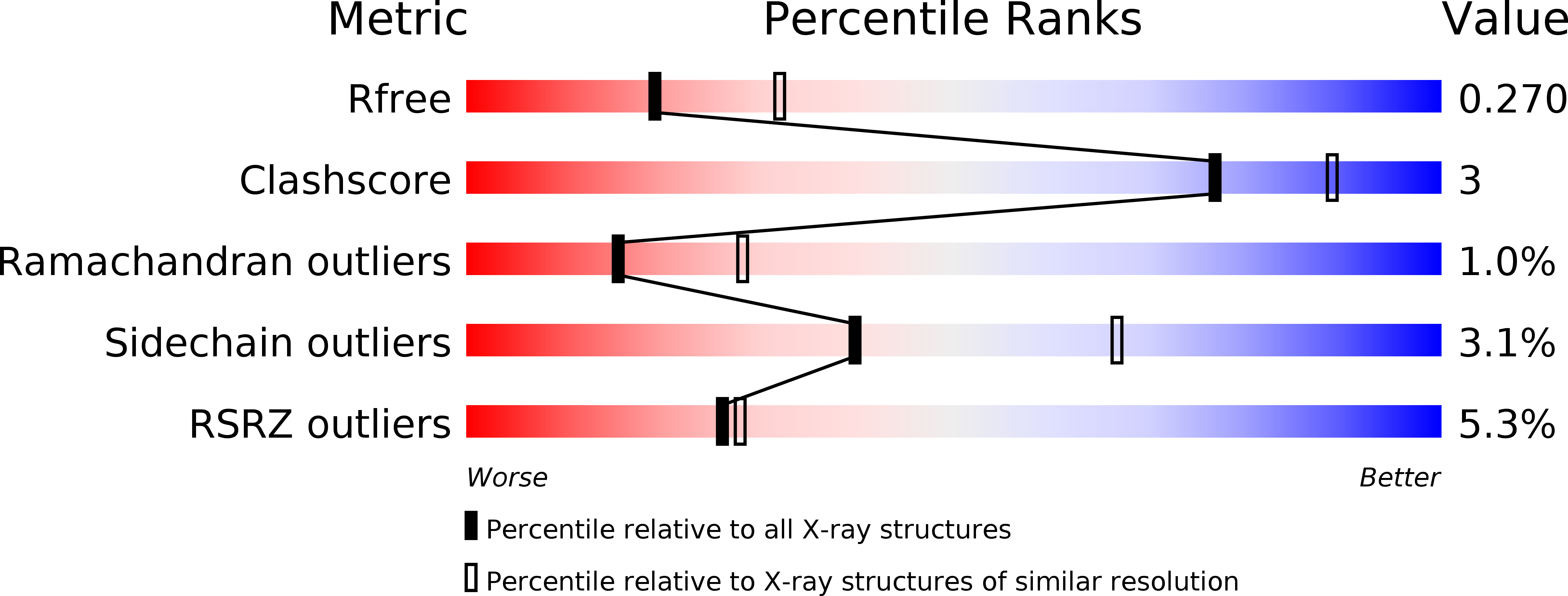
R-Value Free:
0.27
R-Value Work:
0.22
R-Value Observed:
0.23
Space Group:
P 32 2 1