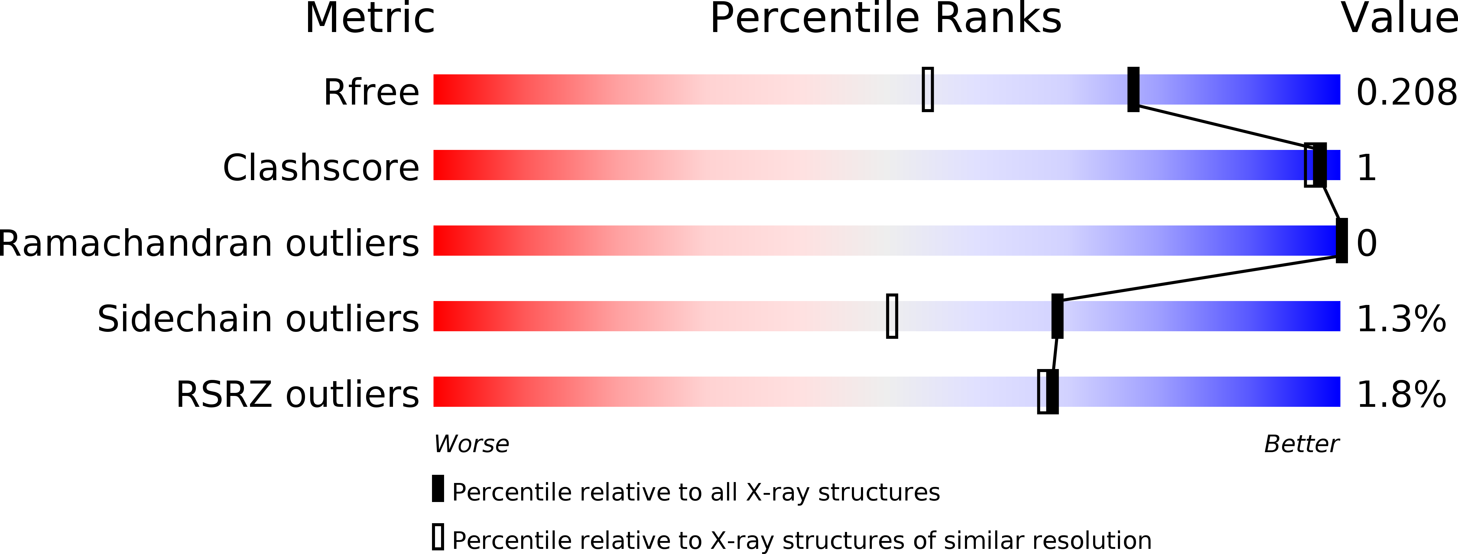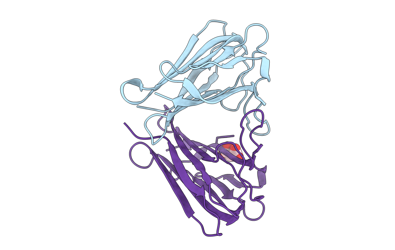
6SVK image
Deposition Date 2019-09-18
Release Date 2019-11-27
Last Version Date 2024-11-13
Entry Detail
PDB ID:
6SVK
Keywords:
Title:
human Myeloid-derived growth factor (MYDGF)
Biological Source:
Source Organism:
Homo sapiens (Taxon ID: 9606)
Host Organism:
Method Details:
Experimental Method:
Resolution:
1.60 Å
R-Value Free:
0.24
R-Value Work:
0.21
R-Value Observed:
0.21
Space Group:
P 31 2 1
Macromolecular Entities
Polymer Type:polypeptide(L)
Molecule:Myeloid-derived growth factor
Gene (Uniprot):MYDGF
Chain IDs:A, B
Chain Length:142
Number of Molecules:2
Biological Source:Homo sapiens
Primary Citation
Crystal structure and receptor-interacting residues of MYDGF - a protein mediating ischemic tissue repair.
Nat Commun 10 5379 5379 (2019)
PMID: 31772377 DOI: 10.1038/s41467-019-13343-7

Abstact

Myeloid-derived growth factor (MYDGF) is a paracrine-acting protein that is produced by bone marrow-derived monocytes and macrophages to protect and repair the heart after myocardial infarction (MI). This effect can be used for the development of protein-based therapies for ischemic tissue repair, also beyond the sole application in heart tissue. Here, we report the X-ray structure of MYDGF and identify its functionally relevant receptor binding epitope. MYDGF consists of a 10-stranded β-sandwich with a folding topology showing no similarities to other cytokines or growth factors. By characterizing the epitope of a neutralizing antibody and utilizing functional assays to study the activity of surface patch-mutations, we were able to localize the receptor interaction interface to a region around two surface tyrosine residues 71 and 73 and an adjacent prominent loop structure of residues 97-101. These findings enable structure-guided protein engineering to develop modified MYDGF variants with potentially improved properties for clinical use.

Legend

Protein

Chemical

Disease

Primary Citation of related structures