6SVF image
Deposition Date 2019-09-18
Release Date 2019-11-20
Last Version Date 2024-01-24
Entry Detail
PDB ID:
6SVF
Title:
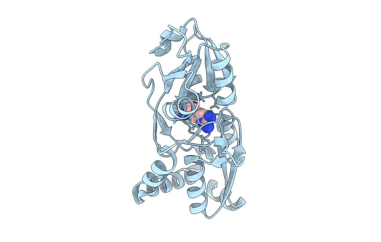
Crystal structure of the P235GK mutant of ArgBP from T. maritima
Biological Source:
Method Details:
Experimental Method:
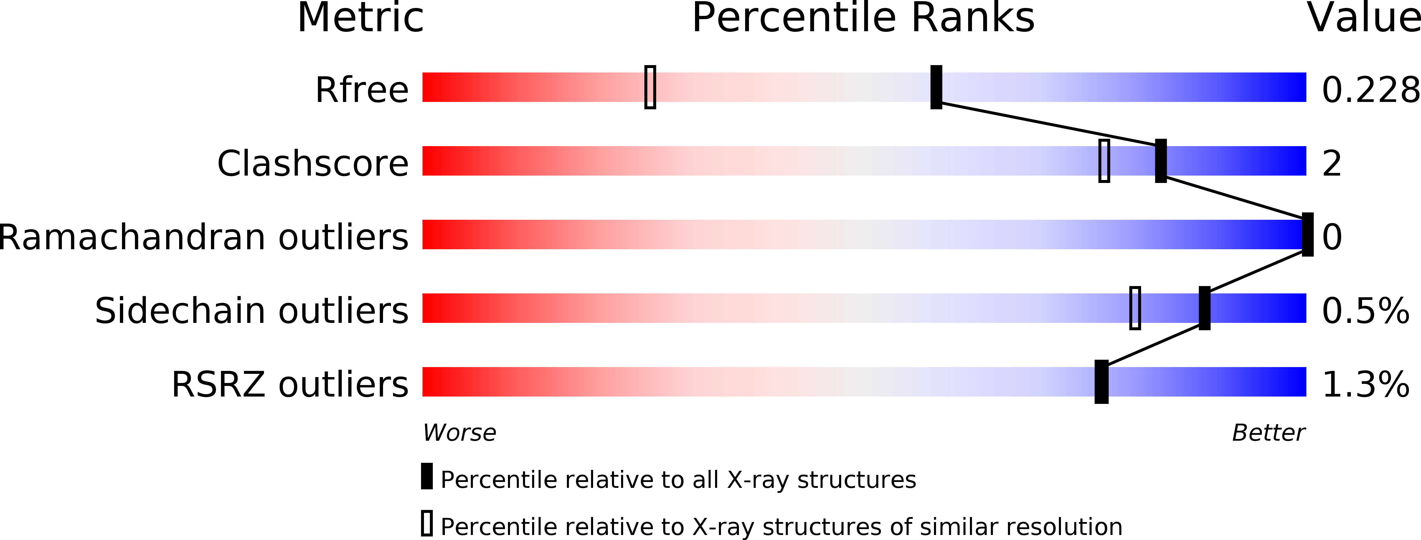
Resolution:
1.60 Å
R-Value Free:
0.21
R-Value Work:
0.17
R-Value Observed:
0.17
Space Group:
P 21 21 21
Macromolecular Entities
Structures with similar UniProt ID
Protein Blast
Polymer Type:polypeptide(L)
Molecule:Amino acid ABC transporter, periplasmic amino acid-binding protein
Gene (Uniprot):TM_0593
Chain IDs:A
Chain Length:229
Number of Molecules:1
Biological Source:Thermotoga maritima (strain ATCC 43589 / MSB8 / DSM 3109 / JCM 10099)
Ligand Molecules
Primary Citation
The non-swapped monomeric structure of the arginine-binding protein from Thermotoga maritima.
Acta Crystallogr.,Sect.F 75 707 713 (2019)
PMID: 31702584 DOI: 10.1107/S2053230X1901464X

Abstact

Domain swapping is a widespread oligomerization process that is observed in a large variety of protein families. In the large superfamily of substrate-binding proteins, non-monomeric members have rarely been reported. The arginine-binding protein from Thermotoga maritima (TmArgBP), a protein endowed with a number of unusual properties, presents a domain-swapped structure in its dimeric native state in which the two polypeptide chains mutually exchange their C-terminal helices. It has previously been shown that mutations in the region connecting the last two helices of the TmArgBP structure lead to the formation of a variety of oligomeric states (monomers, dimers, trimers and larger aggregates). With the aim of defining the structural determinants of domain swapping in TmArgBP, the monomeric form of the P235GK mutant has been structurally characterized. Analysis of this arginine-bound structure indicates that it consists of a closed monomer with its C-terminal helix folded against the rest of the protein, as typically observed for substrate-binding proteins. Notably, the two terminal helices are joined by a single nonhelical residue (Gly235). Collectively, the present findings indicate that extending the hinge region and conferring it with more conformational freedom makes the formation of a closed TmArgBP monomer possible. On the other hand, the short connection between the helices may explain the tendency of the protein to also adopt alternative oligomeric states (dimers, trimers and larger aggregates). The data reported here highlight the importance of evolutionary control to avoid the uncontrolled formation of heterogeneous and potentially harmful oligomeric species through domain swapping.

Legend

Protein

Chemical

Disease

Primary Citation of related structures