Deposition Date
2019-09-18
Release Date
2020-01-22
Last Version Date
2024-11-13
Entry Detail
PDB ID:
6SVB
Keywords:
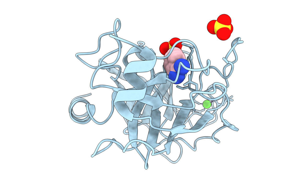
Title:
Terahertz irradiated structure of bovine trypsin (odd frames of crystal x40)
Biological Source:
Source Organism(s):
Bos taurus (Taxon ID: 9913)
Method Details:
Experimental Method:
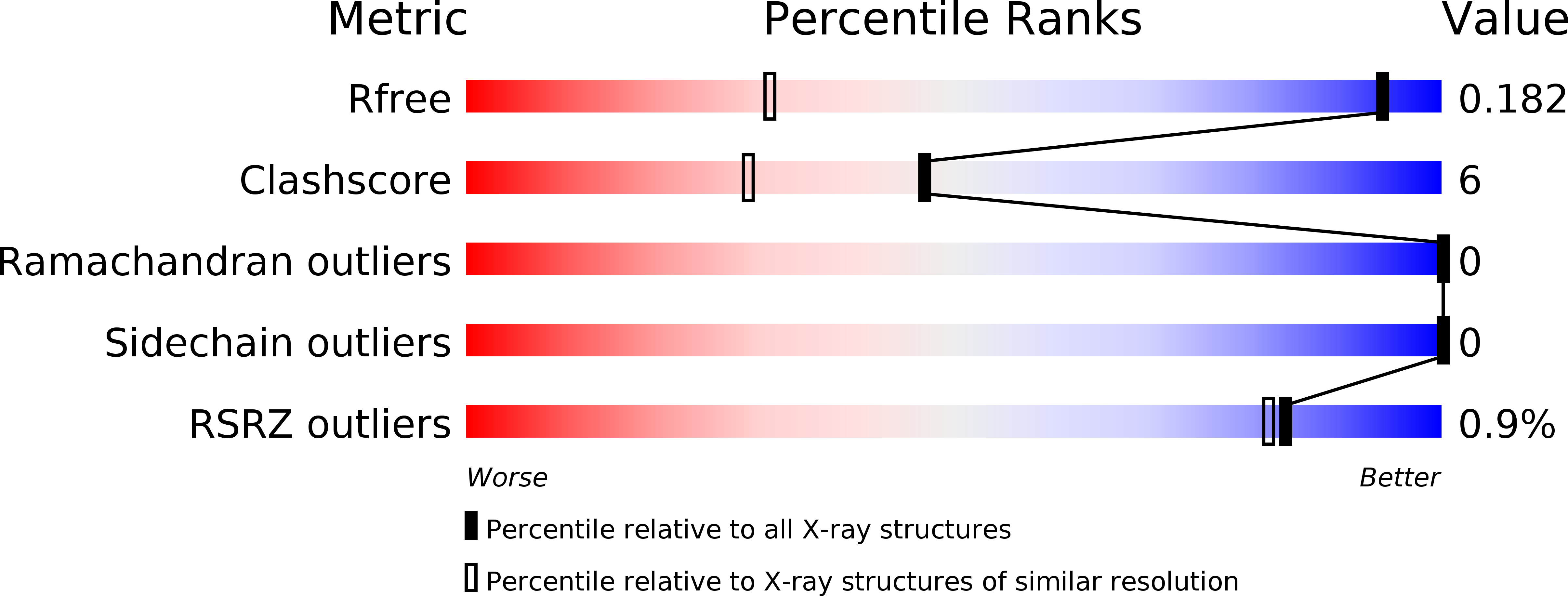
Resolution:
1.15 Å
R-Value Free:
0.18
R-Value Work:
0.13
Space Group:
P 21 21 21