
Deposition Date
2019-09-13
Release Date
2019-11-06
Last Version Date
2024-05-22
Entry Detail
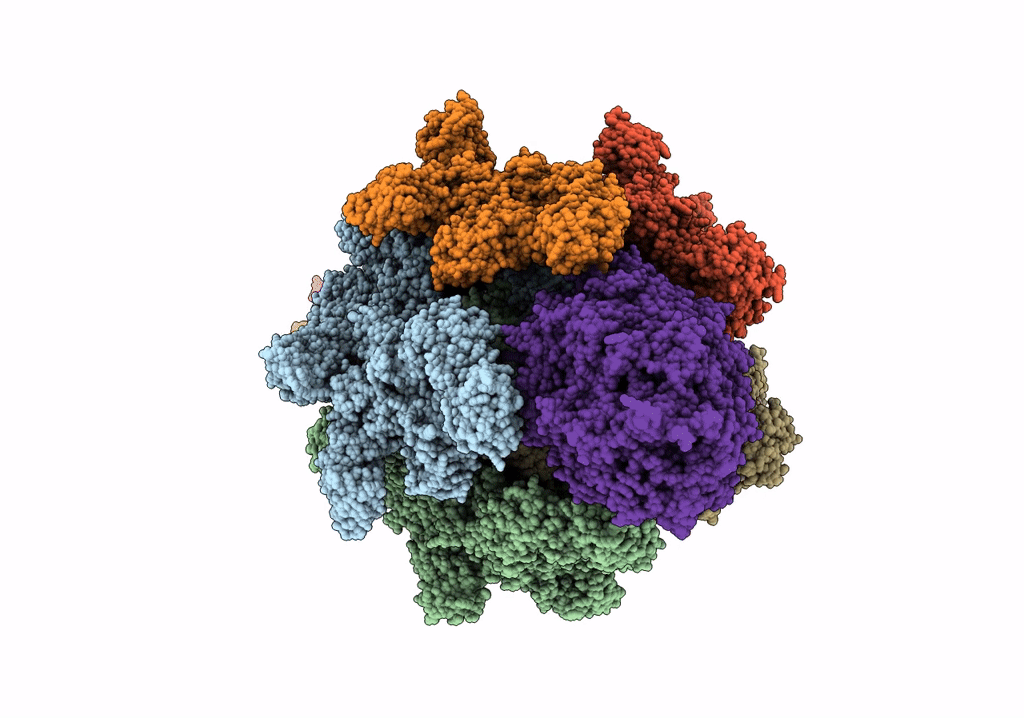
PDB ID:
6SUE
Keywords:
Title:
Structure of Photorhabdus luminescens Tc holotoxin pore, Mutation TccC3-D651A
Biological Source:
Source Organism(s):
Photorhabdus luminescens (Taxon ID: 29488)
Expression System(s):
Method Details:
Experimental Method:
Resolution:
3.40 Å
Aggregation State:
PARTICLE
Reconstruction Method:
SINGLE PARTICLE
