6SU3 image
Entry Detail
PDB ID:
6SU3
Title:
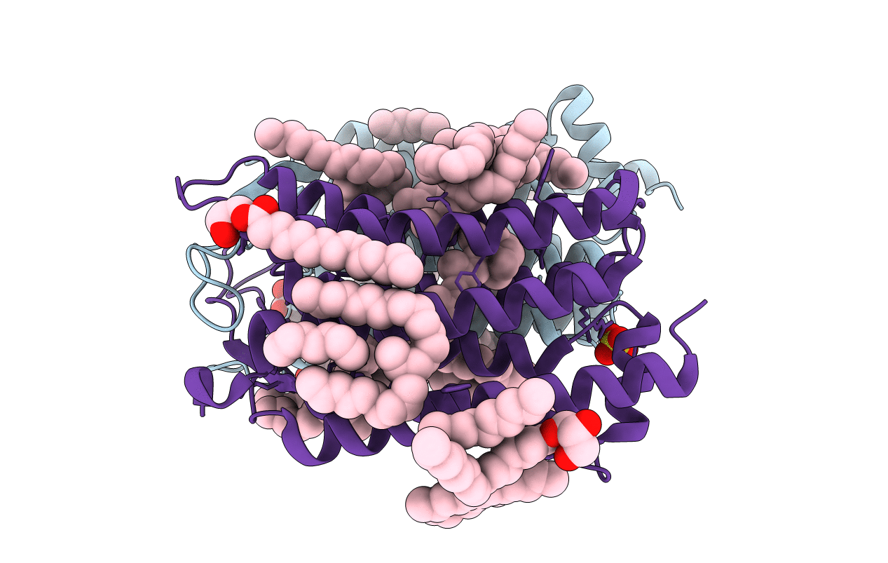
Crystal structure of the 48C12 heliorhodopsin in the violet form at pH 8.8
Biological Source:
Source Organism:
Host Organism:
PDB Version:
Deposition Date:
2019-09-12
Release Date:
2019-12-11
Method Details:
Experimental Method:
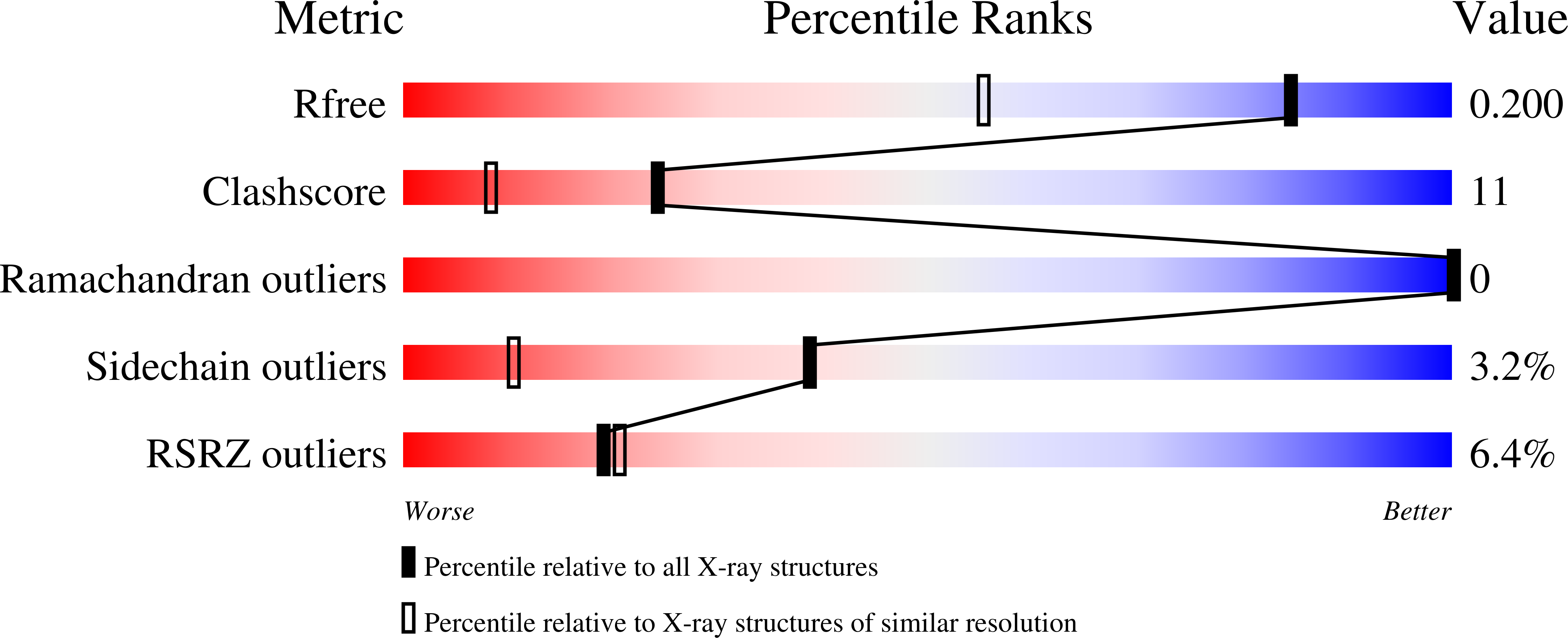
Resolution:
1.50 Å
R-Value Free:
0.19
R-Value Work:
0.15
R-Value Observed:
0.15
Space Group:
P 1 21 1
Macromolecular Entities
Polymer Type:polypeptide(L)
Description:48C12 heliorhodopsin
Chain IDs:A (auth: X), B (auth: A)
Chain Length:264
Number of Molecules:2
Biological Source:Actinobacteria bacterium
Primary Citation
High-resolution structural insights into the heliorhodopsin family.
Proc.Natl.Acad.Sci.USA 117 4131 4141 (2020)
PMID: 32034096 DOI: 10.1073/pnas.1915888117

Abstact

Rhodopsins are the most abundant light-harvesting proteins. A new family of rhodopsins, heliorhodopsins (HeRs), has recently been discovered. Unlike in the known rhodopsins, in HeRs the N termini face the cytoplasm. The function of HeRs remains unknown. We present the structures of the bacterial HeR-48C12 in two states at the resolution of 1.5 Å, which highlight its remarkable difference from all known rhodopsins. The interior of HeR's extracellular part is completely hydrophobic, while the cytoplasmic part comprises a cavity (Schiff base cavity [SBC]) surrounded by charged amino acids and containing a cluster of water molecules, presumably being a primary proton acceptor from the Schiff base. At acidic pH, a planar triangular molecule (acetate) is present in the SBC. Structure-based bioinformatic analysis identified 10 subfamilies of HeRs, suggesting their diverse biological functions. The structures and available data suggest an enzymatic activity of HeR-48C12 subfamily and their possible involvement in fundamental redox biological processes.

Legend

Protein

Chemical

Disease

Primary Citation of related structures