Deposition Date
2019-09-06
Release Date
2021-02-03
Last Version Date
2024-10-23
Entry Detail
PDB ID:
6SRU
Keywords:
Title:
Structure of Ig-like V-type domian of mouse Programmed cell death 1 ligand 1 (PD-L1)
Biological Source:
Source Organism(s):
Mus musculus (Taxon ID: 10090)
Expression System(s):
Method Details:
Experimental Method:
Resolution:
2.53 Å
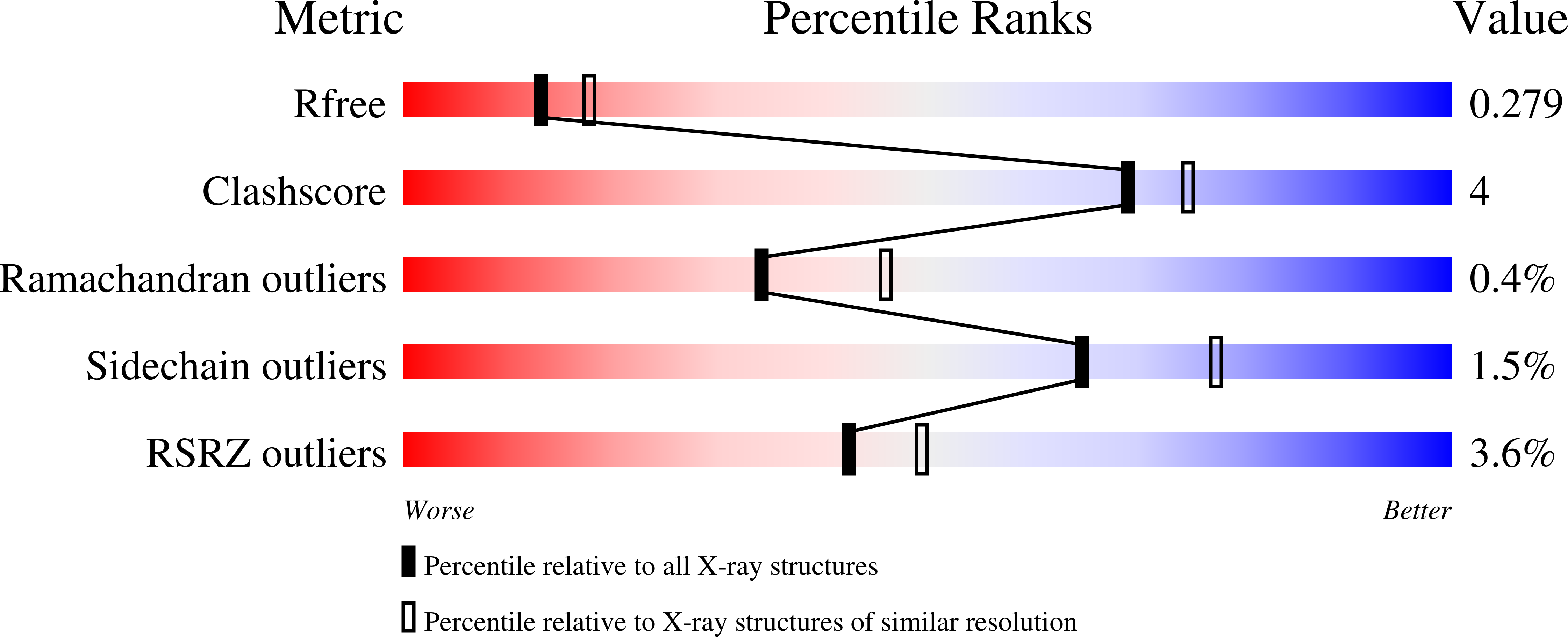
R-Value Free:
0.27
R-Value Work:
0.22
R-Value Observed:
0.23
Space Group:
P 1 21 1