6SMK image
Deposition Date 2019-08-22
Release Date 2020-09-09
Last Version Date 2024-01-31
Entry Detail
PDB ID:
6SMK
Title:
Crystal structure of catalytic domain A109H mutant of prophage-encoded M23 protein EnpA from Enterococcus faecalis.
Biological Source:
Host Organism:
Method Details:
Experimental Method:
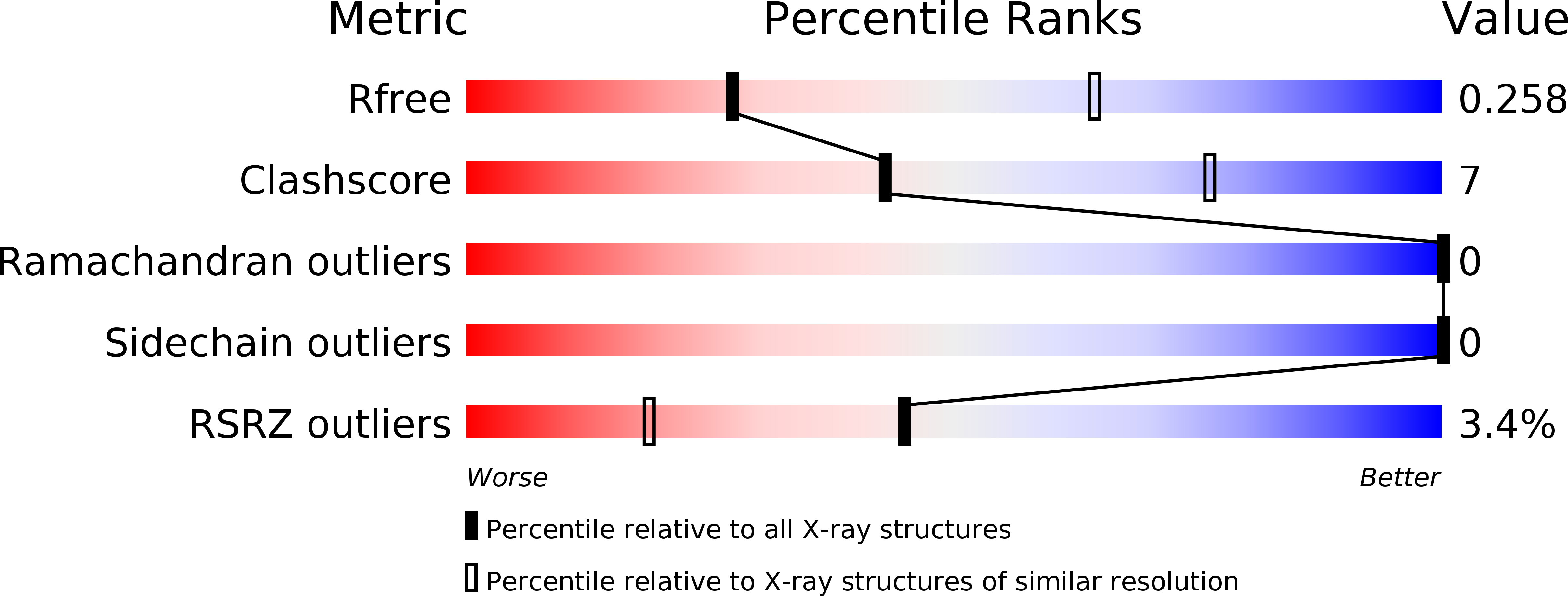
Resolution:
3.00 Å
R-Value Free:
0.25
R-Value Work:
0.22
R-Value Observed:
0.22
Space Group:
P 65 2 2
Macromolecular Entities
Structures with similar UniProt ID
Protein Blast
Polymer Type:polypeptide(L)
Molecule:Peptidase_M23 domain-containing protein
Gene (Uniprot):EF_1473
Mutagens:A109H
Chain IDs:A, B, C (auth: D), D (auth: C), E
Chain Length:142
Number of Molecules:5
Biological Source:Enterococcus faecalis (strain ATCC 700802 / V583)
Ligand Molecules
Primary Citation
Structural Characterization of EnpA D,L-Endopeptidase from Enterococcus faecalis Prophage Provides Insights into Substrate Specificity of M23 Peptidases.
Int J Mol Sci 22 ? ? (2021)
PMID: 34281200 DOI: 10.3390/ijms22137136

Abstact

The best-characterized members of the M23 family are glycyl-glycine hydrolases, such as lysostaphin (Lss) from Staphylococcus simulans or LytM from Staphylococcus aureus. Recently, enzymes with broad specificities were reported, such as EnpACD from Enterococcus faecalis, that cleaves D,L peptide bond between the stem peptide and a cross-bridge. Previously, the activity of EnpACD was demonstrated only on isolated peptidoglycan fragments. Herein we report conditions in which EnpACD lyses bacterial cells live with very high efficiency demonstrating great bacteriolytic potential, though limited to a low ionic strength environment. We have solved the structure of the EnpACD H109A inactive variant and analyzed it in the context of related peptidoglycan hydrolases structures to reveal the bases for the specificity determination. All M23 structures share a very conserved β-sheet core which constitutes the rigid bottom of the substrate-binding groove and active site, while variable loops create the walls of the deep and narrow binding cleft. A detailed analysis of the binding groove architecture, specificity of M23 enzymes and D,L peptidases demonstrates that the substrate groove, which is particularly deep and narrow, is accessible preferably for peptides composed of amino acids with short side chains or subsequent L and D-isomers. As a result, the bottom of the groove is involved in interactions with the main chain of the substrate while the side chains are protruding in one plane towards the groove opening. We concluded that the selectivity of the substrates is based on their conformations allowed only for polyglycine chains and alternating chirality of the amino acids.

Legend

Protein

Chemical

Disease

Primary Citation of related structures
Feedback Form
Name
Email
Institute
Feedback