Deposition Date
2019-08-11
Release Date
2019-12-18
Last Version Date
2024-01-24
Entry Detail
PDB ID:
6SIT
Keywords:
Title:
Pseudo-atomic crystal structure of the desmoglein 2 - human adenovirus serotype 3 fibre knob complex
Biological Source:
Source Organism(s):
Human adenovirus B3 (Taxon ID: 45659)
Homo sapiens (Taxon ID: 9606)
Homo sapiens (Taxon ID: 9606)
Expression System(s):
Method Details:
Experimental Method:
Resolution:
4.50 Å
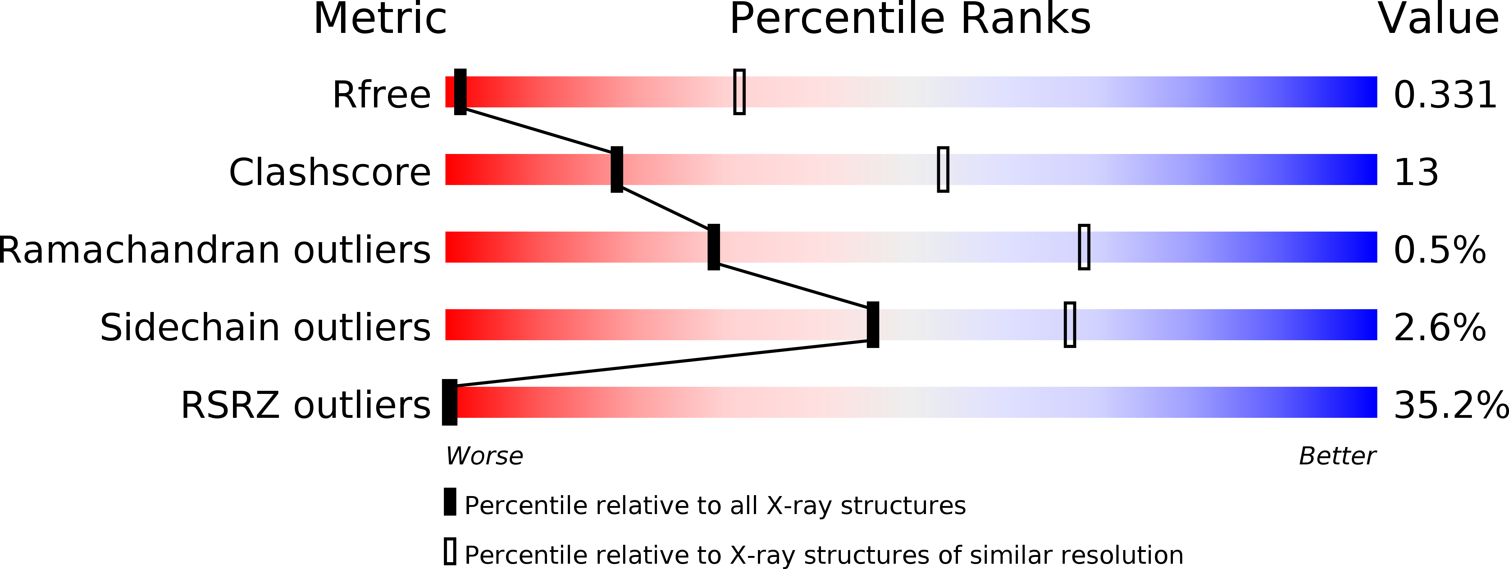
R-Value Free:
0.37
R-Value Work:
0.36
Space Group:
I 21 3