Deposition Date
2019-07-28
Release Date
2019-10-23
Last Version Date
2024-11-06
Entry Detail
PDB ID:
6SDK
Keywords:
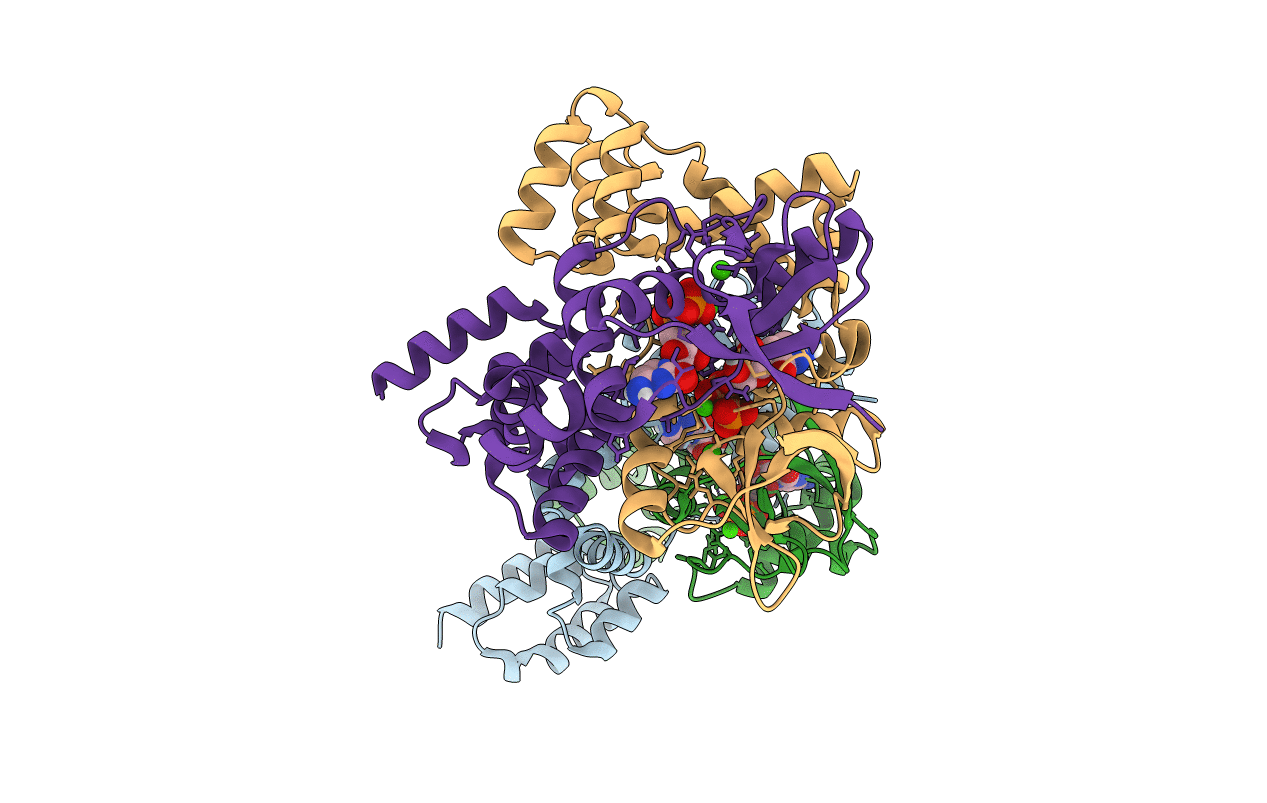
Title:
Crystal structure of bacterial ParB dimer bound to CDP
Biological Source:
Source Organism(s):
Bacillus subtilis (strain 168) (Taxon ID: 224308)
Expression System(s):
Method Details:
Experimental Method:
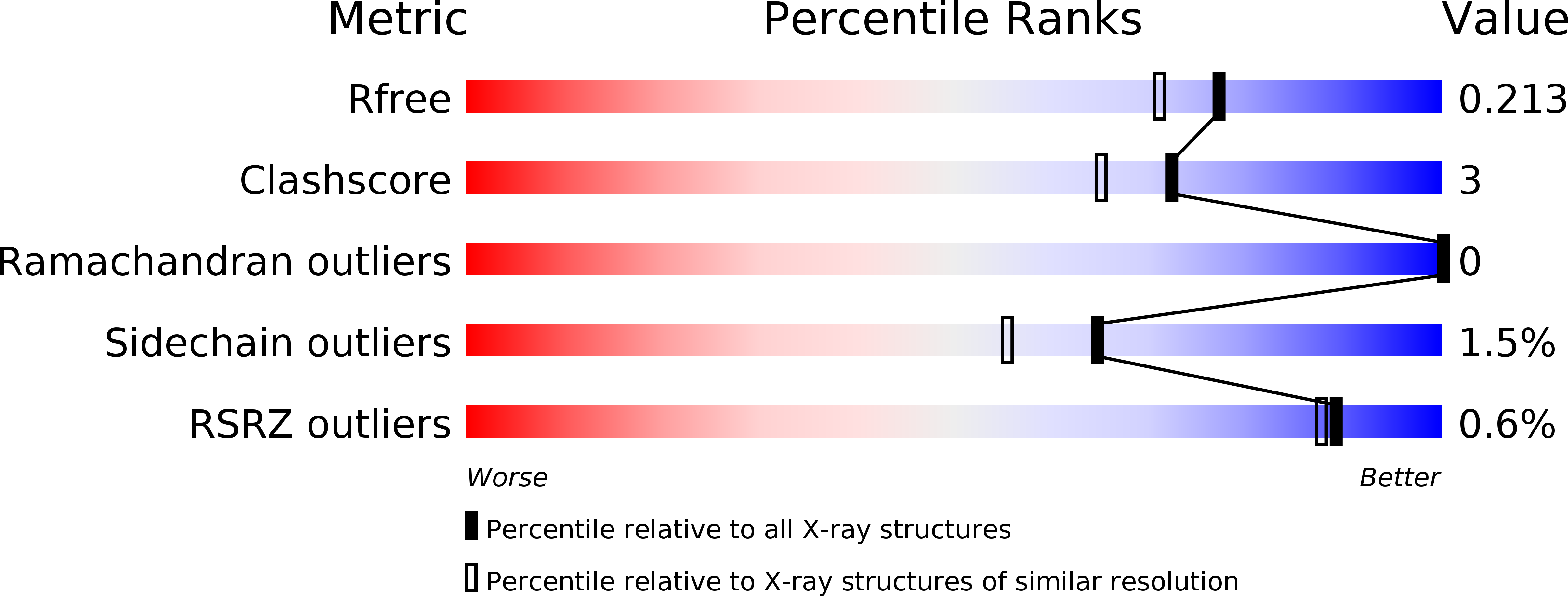
Resolution:
1.81 Å
R-Value Free:
0.21
R-Value Work:
0.18
R-Value Observed:
0.18
Space Group:
P 1 21 1