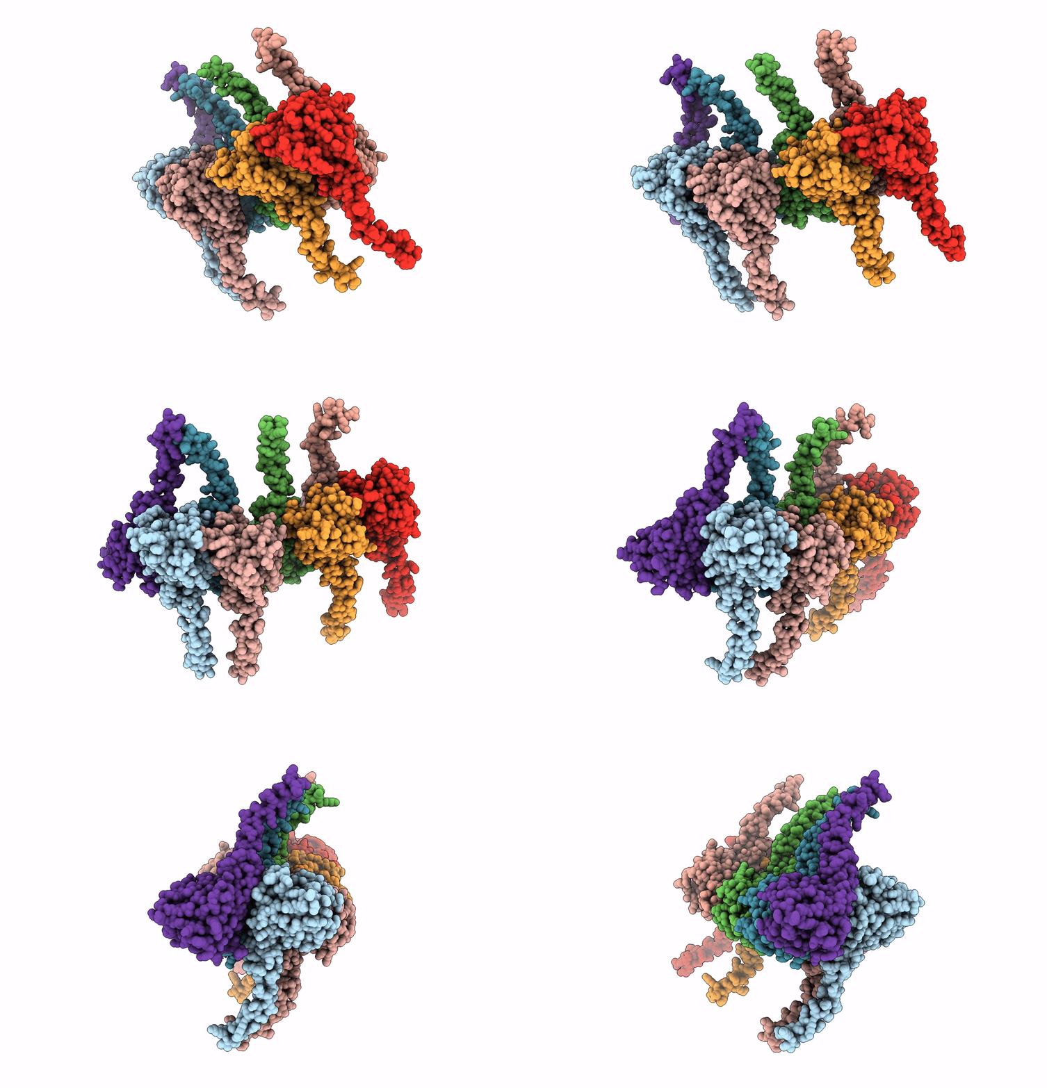
6SB4 image
Entry Detail
PDB ID:
6SB4
Keywords:
Title:
Crystal structure of murine perforin-2 P2 domain crystal form 2
Biological Source:
Source Organism:
Host Organism:
PDB Version:
Deposition Date:
2019-07-18
Release Date:
2020-02-05
Method Details:
Experimental Method:
Resolution:
3.17 Å
R-Value Free:
0.22
R-Value Work:
0.18
R-Value Observed:
0.19
Space Group:
P 1
Macromolecular Entities
Polymer Type:polypeptide(L)
Description:Macrophage-expressed gene 1 protein
Chain IDs:A, B, C, D, E, F, G, H
Chain Length:295
Number of Molecules:8
Biological Source:Mus musculus
Ligand Molecules
Primary Citation
Structure and mechanism of bactericidal mammalian perforin-2, an ancient agent of innate immunity.
Sci Adv 6 eaax8286 eaax8286 (2020)
PMID: 32064340 DOI: 10.1126/sciadv.aax8286

Abstact

Perforin-2 (MPEG1) is thought to enable the killing of invading microbes engulfed by macrophages and other phagocytes, forming pores in their membranes. Loss of perforin-2 renders individual phagocytes and whole organisms significantly more susceptible to bacterial pathogens. Here, we reveal the mechanism of perforin-2 activation and activity using atomic structures of pre-pore and pore assemblies, high-speed atomic force microscopy, and functional assays. Perforin-2 forms a pre-pore assembly in which its pore-forming domain points in the opposite direction to its membrane-targeting domain. Acidification then triggers pore formation, via a 180° conformational change. This novel and unexpected mechanism prevents premature bactericidal attack and may have played a key role in the evolution of all perforin family proteins.

Legend

Protein

Chemical

Disease

Primary Citation of related structures