6S9K image
Entry Detail
PDB ID:
6S9K
Title:
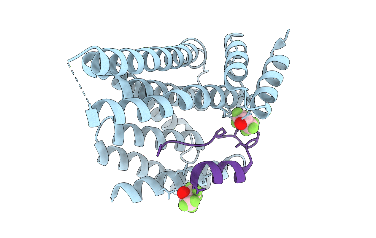
Structure of 14-3-3 gamma in complex with caspase-2 peptide containing 14-3-3 binding motif Ser139 and NLS
Biological Source:
Source Organism:
PDB Version:
Deposition Date:
2019-07-15
Release Date:
2020-01-22
Method Details:
Experimental Method:
Resolution:
1.60 Å
R-Value Free:
0.22
R-Value Work:
0.20
R-Value Observed:
0.20
Space Group:
I 2 2 2
Macromolecular Entities
Polymer Type:polypeptide(L)
Description:14-3-3 protein gamma
Mutations:none
Chain IDs:A
Chain Length:234
Number of Molecules:1
Biological Source:Homo sapiens
Polymer Type:polypeptide(L)
Description:Caspase-2
Chain IDs:B
Chain Length:34
Number of Molecules:1
Biological Source:Homo sapiens
Modified Residue
Compound ID Chain ID Parent Comp ID Details 2D Image
SEP B SER modified residue
Ligand Molecules
Primary Citation
14-3-3 protein binding blocks the dimerization interface of caspase-2.
Febs J. 287 3494 3510 (2020)
PMID: 31961068 DOI: 10.1111/febs.15215

Abstact

Among all species, caspase-2 (C2) is the most evolutionarily conserved caspase required for effective initiation of apoptosis following death stimuli. C2 is activated through dimerization and autoproteolytic cleavage and inhibited through phosphorylation at Ser139 and Ser164 , within the linker between the caspase recruitment and p19 domains of the zymogen, followed by association with the adaptor protein 14-3-3, which maintains C2 in its immature form procaspase (proC2). However, the mechanism of 14-3-3-dependent inhibition of C2 activation remains unclear. Here, we report the structural characterization of the complex between proC2 and 14-3-3 by hydrogen/deuterium mass spectrometry and protein crystallography to determine the molecular basis for 14-3-3-mediated inhibition of C2 activation. Our data reveal that the 14-3-3 dimer interacts with proC2 not only through ligand-binding grooves but also through other regions outside the central channel, thus explaining the isoform-dependent specificity of 14-3-3 protein binding to proC2 and the substantially higher binding affinity of 14-3-3 protein to proC2 than to the doubly phosphorylated peptide. The formation of the complex between 14-3-3 protein and proC2 does not induce any large conformational change in proC2. Furthermore, 14-3-3 protein interacts with and masks both the nuclear localization sequence and the C-terminal region of the p12 domain of proC2 through transient interactions in which both the p19 and p12 domains of proC2 are not firmly docked onto the surface of 14-3-3. This masked region of p12 domain is involved in C2 dimerization. Therefore, 14-3-3 protein likely inhibits proC2 activation by blocking its dimerization surface. DATABASES: Structural data are available in the Protein Data Bank under the accession numbers 6SAD and 6S9K.

Legend

Protein

Chemical

Disease

Primary Citation of related structures