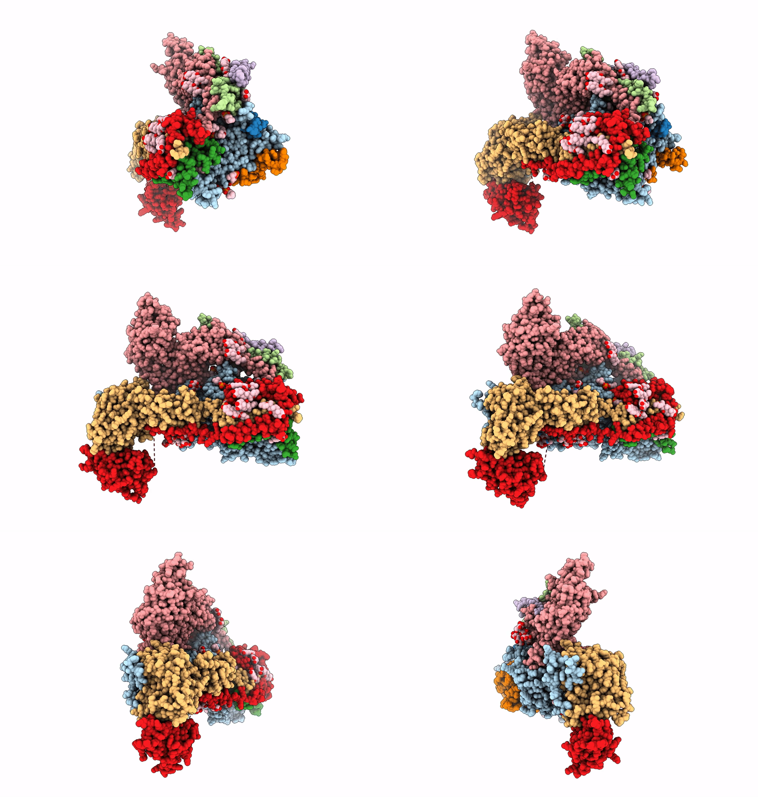
6S7T image
Entry Detail
PDB ID:
6S7T
EMDB ID:
Keywords:
Title:
Cryo-EM structure of human oligosaccharyltransferase complex OST-B
Biological Source:
Source Organism:
Host Organism:
PDB Version:
Deposition Date:
2019-07-05
Release Date:
2019-12-18
Method Details:
Experimental Method:
Resolution:
3.50 Å
Aggregation State:
PARTICLE
Reconstruction Method:
SINGLE PARTICLE
Macromolecular Entities
Polymer Type:polypeptide(L)
Description:Dolichyl-diphosphooligosaccharide--protein glycosyltransferase subunit STT3B
Chain IDs:A
Chain Length:826
Number of Molecules:1
Biological Source:Homo sapiens
Polymer Type:polypeptide(L)
Description:Dolichyl-diphosphooligosaccharide--protein glycosyltransferase subunit 4
Chain IDs:B
Chain Length:37
Number of Molecules:1
Biological Source:Homo sapiens
Polymer Type:polypeptide(L)
Description:Transmembrane protein 258
Chain IDs:C
Chain Length:79
Number of Molecules:1
Biological Source:Homo sapiens
Polymer Type:polypeptide(L)
Description:Dolichyl-diphosphooligosaccharide--protein glycosyltransferase subunit DAD1
Chain IDs:D
Chain Length:113
Number of Molecules:1
Biological Source:Homo sapiens
Polymer Type:polypeptide(L)
Description:Dolichyl-diphosphooligosaccharide--protein glycosyltransferase subunit 1
Chain IDs:E
Chain Length:607
Number of Molecules:1
Biological Source:Homo sapiens
Polymer Type:polypeptide(L)
Description:Dolichyl-diphosphooligosaccharide--protein glycosyltransferase subunit 2
Chain IDs:F
Chain Length:631
Number of Molecules:1
Biological Source:Homo sapiens
Polymer Type:polypeptide(L)
Description:Dolichyl-diphosphooligosaccharide--protein glycosyltransferase 48 kDa subunit
Chain IDs:G
Chain Length:456
Number of Molecules:1
Biological Source:Homo sapiens
Polymer Type:polypeptide(L)
Description:Magnesium transporter protein 1
Chain IDs:H
Chain Length:335
Number of Molecules:1
Biological Source:Homo sapiens
Polymer Type:polypeptide(L)
Description:Malectin
Chain IDs:I
Chain Length:292
Number of Molecules:1
Biological Source:Homo sapiens
Polymer Type:polypeptide(L)
Description:PEPTIDE
Chain IDs:J (auth: K)
Chain Length:7
Number of Molecules:1
Biological Source:Homo sapiens
Primary Citation
Cryo-electron microscopy structures of human oligosaccharyltransferase complexes OST-A and OST-B.
Science 366 1372 1375 (2019)
PMID: 31831667 DOI: 10.1126/science.aaz3505

Abstact

Oligosaccharyltransferase (OST) catalyzes the transfer of a high-mannose glycan onto secretory proteins in the endoplasmic reticulum. Mammals express two distinct OST complexes that act in a cotranslational (OST-A) or posttranslocational (OST-B) manner. Here, we present high-resolution cryo-electron microscopy structures of human OST-A and OST-B. Although they have similar overall architectures, structural differences in the catalytic subunits STT3A and STT3B facilitate contacts to distinct OST subunits, DC2 in OST-A and MAGT1 in OST-B. In OST-A, interactions with TMEM258 and STT3A allow ribophorin-I to form a four-helix bundle that can bind to a translating ribosome, whereas the equivalent region is disordered in OST-B. We observed an acceptor peptide and dolichylphosphate bound to STT3B, but only dolichylphosphate in STT3A, suggesting distinct affinities of the two OST complexes for protein substrates.

Legend

Protein

Chemical

Disease

Primary Citation of related structures