6RWD image
Deposition Date 2019-06-04
Release Date 2019-06-19
Last Version Date 2024-01-24
Entry Detail
PDB ID:
6RWD
Keywords:
Title:
Crystal structure of SjGST in complex with GSH and ellagic acid at 1.53 Angstrom resolution
Biological Source:
Source Organism:
Host Organism:
Method Details:
Experimental Method:
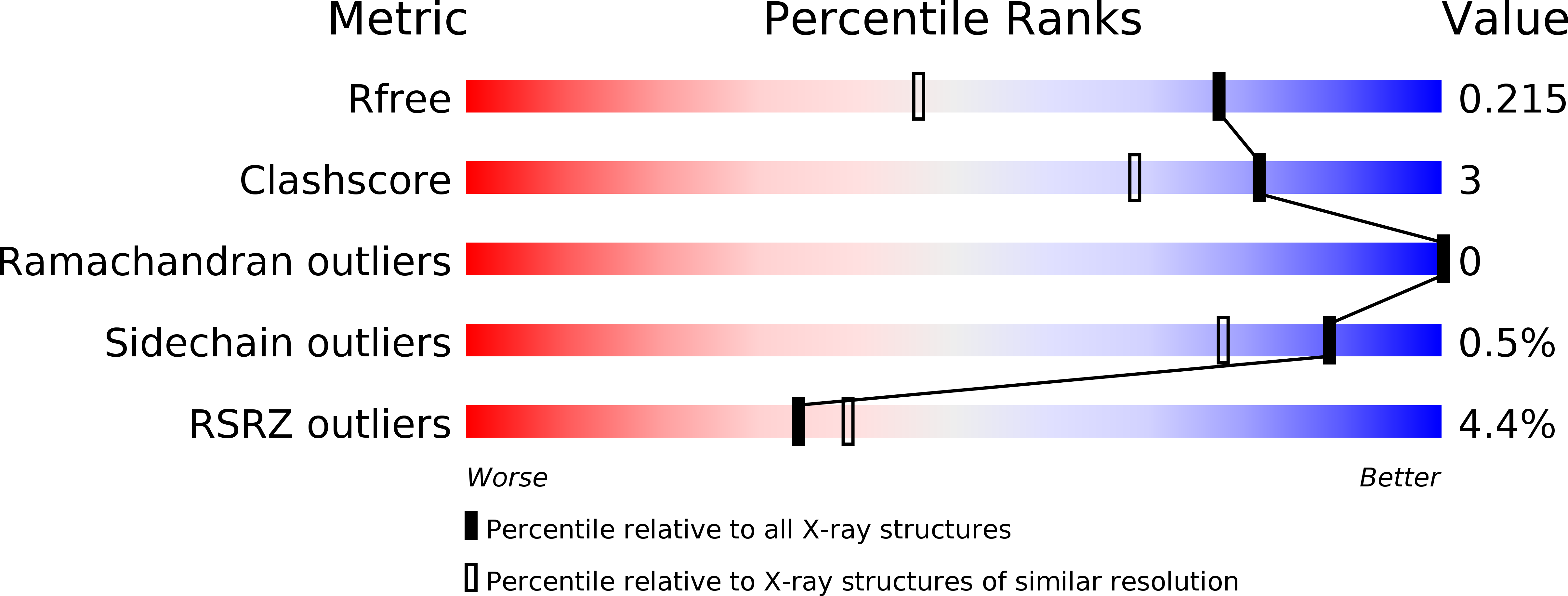
Resolution:
1.53 Å
R-Value Free:
0.21
R-Value Work:
0.18
R-Value Observed:
0.18
Space Group:
P 21 21 21
Macromolecular Entities
Structures with similar UniProt ID
Protein Blast
Polymer Type:polypeptide(L)
Molecule:Glutathione S-transferase class-mu 26 kDa isozyme
Chain IDs:A, B
Chain Length:218
Number of Molecules:2
Biological Source:Schistosoma japonicum
Primary Citation
Molecular basis of inhibition of Schistosoma japonicum glutathione transferase by ellagic acid: Insights into biophysical and structural studies.
Mol.Biochem.Parasitol. 240 111319 111319 (2020)
PMID: 32961204 DOI: 10.1016/j.molbiopara.2020.111319

Abstact

Schistosoma japonicum glutathione transferase (Sj26GST), an enzyme central to detoxification of electrophilic compounds in the parasite, is upregulated in response to drug treatment. Therefore, Sj26GST may serve as a potential therapeutic target for the treatment of schistosomiasis. Herewith, we describe the structural basis of inhibition of Sj26GST by ellagic acid (EA). Using 1-chloro-2,4-dinitrobenzene and reduced glutathione (GSH) as Sj26GST substrates, EA was shown to inhibit Sj26GST activity by 66 % with an IC50 of 2.4 μM. Fluorescence spectroscopy showed that EA altered the polarity of the environment of intrinsic tryptophan and that EA decreased (in a dose-dependent manner) the interaction between Sj26GST and 8-Anilino-1-naphthalenesulfonate (ANS), which is a known GST H-site ligand. Thermodynamic studies indicated that the interaction between Sj26GST and EA is spontaneous (ΔG = -29.88 ± 0.07 kJ/mol), enthalpically-driven (ΔH = -9.48 ± 0.42 kJ/mol) with a favourable entropic change (ΔS = 20.40 ± 0.08 kJ/mol/K), and with a stoichiometry of four EA molecules bound per Sj26GST dimer. The 1.53 Å-resolution Sj26GST crystal structure (P 21 21 21 space group) complexed with GSH and EA shows that EA binds primarily at the dimer interface, stabilised largely by Van der Waal forces and H-bonding. Besides, EA bound near the H-site and less than 3.5 Å from the ε-NH2 of the γ-glutamyl moiety of GSH, in each subunit.

Legend

Protein

Chemical

Disease

Primary Citation of related structures