6RV7 image
Entry Detail
PDB ID:
6RV7
Keywords:
Title:
Crystal Structure of Glucuronoyl Esterase from Cerrena unicolor inactive S270A variant in complex with the aldouronic acid UXXR
Biological Source:
Source Organism:
Host Organism:
PDB Version:
Deposition Date:
2019-05-31
Release Date:
2020-03-18
Method Details:
Experimental Method:
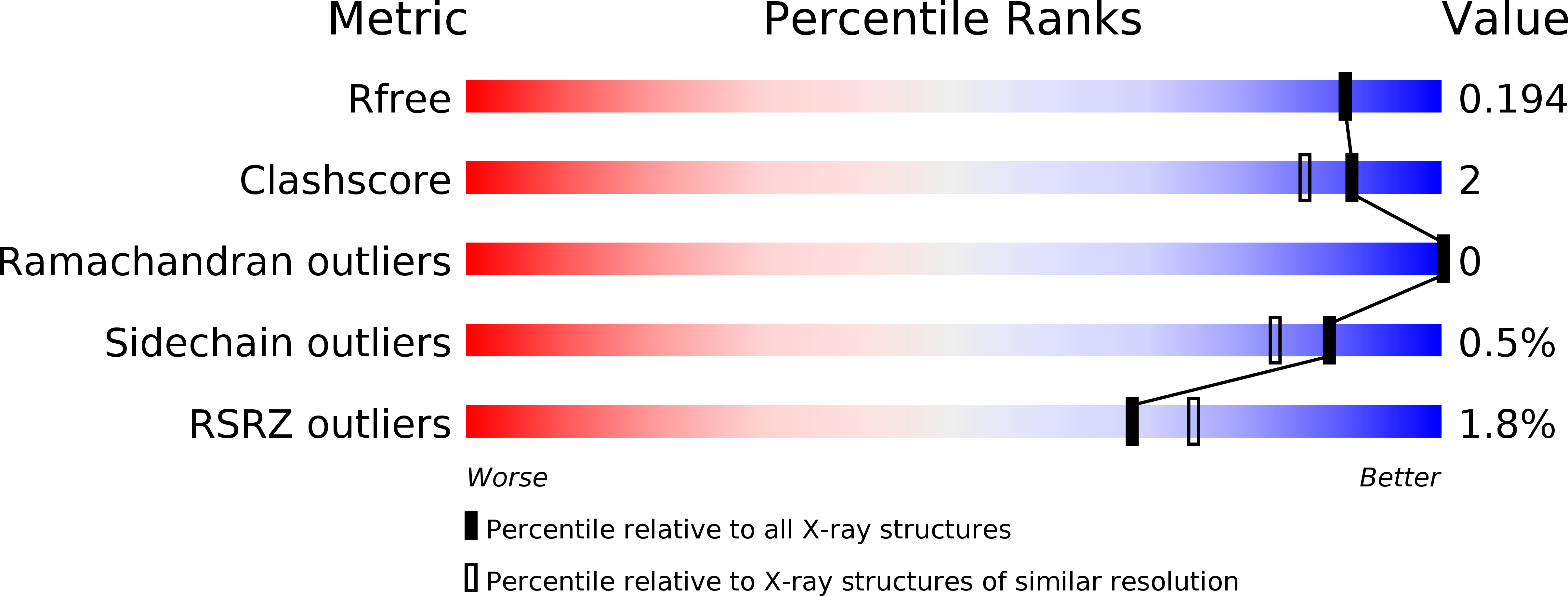
Resolution:
1.73 Å
R-Value Free:
0.19
R-Value Work:
0.16
R-Value Observed:
0.16
Space Group:
P 41 21 2
Macromolecular Entities
Polymer Type:polypeptide(L)
Description:4-O-methyl-glucuronoyl methylesterase
Mutations:S270A
Chain IDs:A, B
Chain Length:401
Number of Molecules:2
Biological Source:Cerrena unicolor
Peptide-like Molecules
PRD_900116
Primary Citation
The structural basis of fungal glucuronoyl esterase activity on natural substrates.
Nat Commun 11 1026 1026 (2020)
PMID: 32094331 DOI: 10.1038/s41467-020-14833-9

Abstact

Structural and functional studies were conducted of the glucuronoyl esterase (GE) from Cerrena unicolor (CuGE), an enzyme catalyzing cleavage of lignin-carbohydrate ester bonds. CuGE is an α/β-hydrolase belonging to carbohydrate esterase family 15 (CE15). The enzyme is modular, comprised of a catalytic and a carbohydrate-binding domain. SAXS data show CuGE as an elongated rigid molecule where the two domains are connected by a rigid linker. Detailed structural information of the catalytic domain in its apo- and inactivated form and complexes with aldouronic acids reveal well-defined binding of the 4-O-methyl-a-D-glucuronoyl moiety, not influenced by the nature of the attached xylo-oligosaccharide. Structural and sequence comparisons within CE15 enzymes reveal two distinct structural subgroups. CuGE belongs to the group of fungal CE15-B enzymes with an open and flat substrate-binding site. The interactions between CuGE and its natural substrates are explained and rationalized by the structural results, microscale thermophoresis and isothermal calorimetry.

Legend

Protein

Chemical

Disease

Primary Citation of related structures