6RV5 image
Deposition Date 2019-05-31
Release Date 2020-04-01
Last Version Date 2024-01-31
Entry Detail
PDB ID:
6RV5
Keywords:
Title:
X-ray structure of the levansucrase from Erwinia tasmaniensis in complex with levanbiose
Biological Source:
Method Details:
Experimental Method:
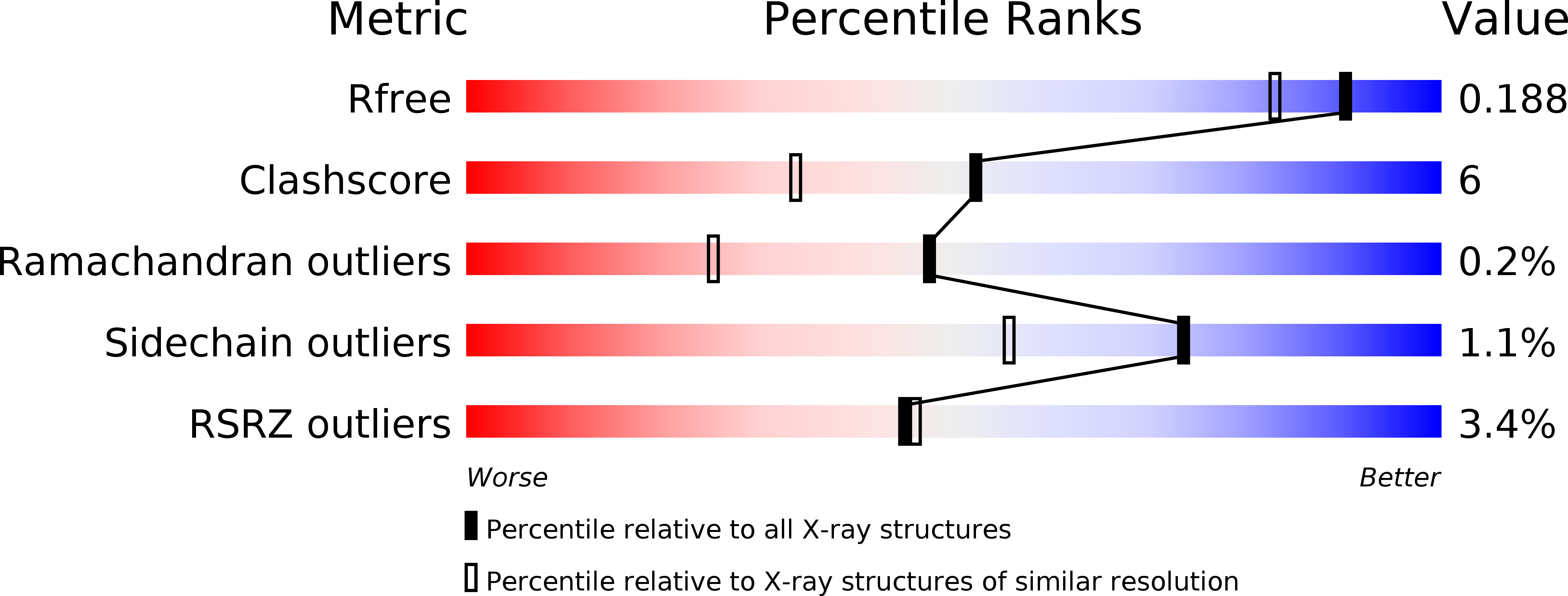
Resolution:
1.58 Å
R-Value Free:
0.19
R-Value Work:
0.14
R-Value Observed:
0.12
Space Group:
P 41 21 2
Macromolecular Entities
Structures with similar UniProt ID
Protein Blast
Polymer Type:polypeptide(L)
Molecule:Levansucrase (Beta-D-fructofuranosyl transferase)
Gene (Uniprot):lsc
Chain IDs:A
Chain Length:412
Number of Molecules:1
Biological Source:Erwinia tasmaniensis (strain DSM 17950 / CIP 109463 / Et1/99)
Peptide-like Molecules
PRD_900051
Primary Citation
The Structure of Sucrose-Soaked Levansucrase Crystals fromErwinia tasmaniensisreveals a Binding Pocket for Levanbiose.
Int J Mol Sci 21 ? ? (2019)
PMID: 31877648 DOI: 10.3390/ijms21010083

Abstact

Given its potential role in the synthesis of novel prebiotics and applications in the pharmaceutical industry, a strong interest has developed in the enzyme levansucrase (LSC, EC 2.4.1.10). LSC catalyzes both the hydrolysis of sucrose (or sucroselike substrates) and the transfructosylation of a wide range of acceptors. LSC from the Gram-negative bacterium Erwinia tasmaniensis (EtLSC) is an interesting biocatalyst due to its high-yield production of fructooligosaccharides (FOSs). In order to learn more about the process of chain elongation, we obtained the crystal structure of EtLSC in complex with levanbiose (LBS). LBS is an FOS intermediate formed during the synthesis of longer-chain FOSs and levan. Analysis of the LBS binding pocket revealed that its structure was conserved in several related species. The binding pocket discovered in this crystal structure is an ideal target for future mutagenesis studies in order to understand its biological relevance and to engineer LSCs into tailored products.

Legend

Protein

Chemical

Disease

Primary Citation of related structures