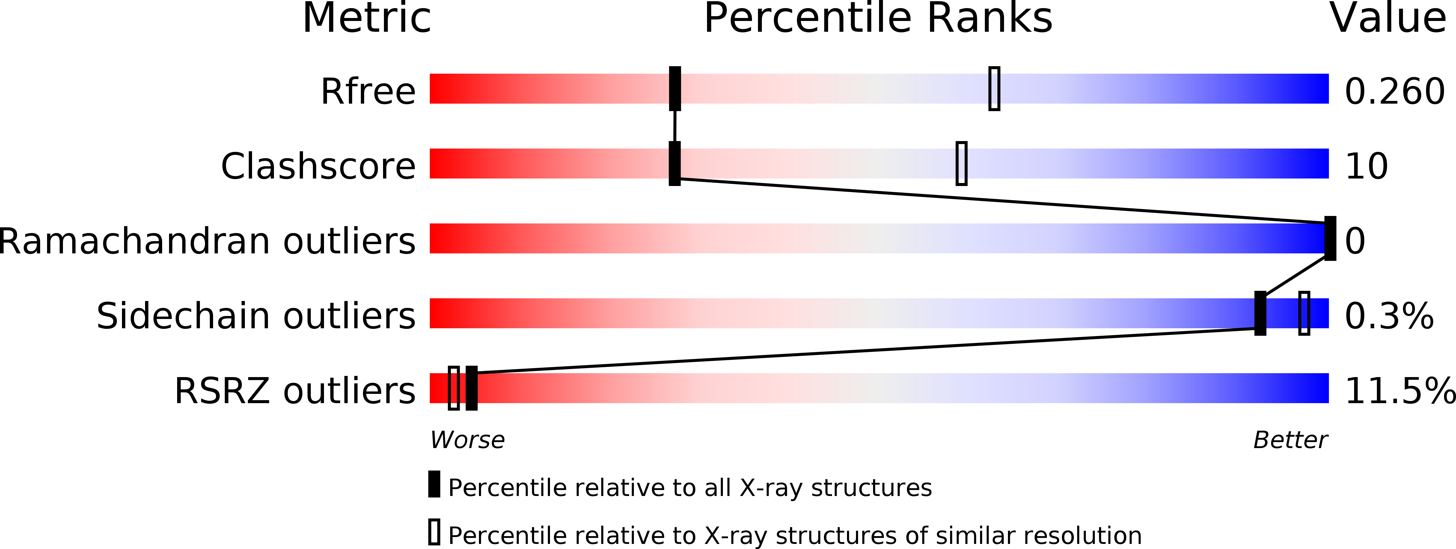
6RUU image
Deposition Date 2019-05-29
Release Date 2020-09-09
Last Version Date 2024-11-20
Entry Detail
PDB ID:
6RUU
Title:
Pseudokinase domain of human IRAK3
Biological Source:
Source Organism:
Homo sapiens (Taxon ID: 9606)
Host Organism:
Method Details:
Experimental Method:
Resolution:
2.95 Å
R-Value Free:
0.26
R-Value Work:
0.22
R-Value Observed:
0.22
Space Group:
P 21 21 2
Macromolecular Entities
Structures with similar UniProt ID
Protein Blast
Polymer Type:polypeptide(L)
Molecule:Interleukin-1 receptor-associated kinase 3
Gene (Uniprot):IRAK3
Chain IDs:A, B, C
Chain Length:315
Number of Molecules:3
Biological Source:Homo sapiens
Primary Citation
Dimeric Structure of the Pseudokinase IRAK3 Suggests an Allosteric Mechanism for Negative Regulation.
Structure 29 238 ? (2021)
PMID: 33238146 DOI: 10.1016/j.str.2020.11.004

Abstact

Interleukin-1 receptor associated kinases (IRAKs) are key players in innate immune signaling that mediate the host response to pathogens. In contrast to the active kinases IRAK1 and IRAK4, IRAK2 and IRAK3 are pseudokinases lacking catalytic activity and their functions are poorly understood. IRAK3 is thought to be a negative regulator of innate immune signaling and mutations in IRAK3 are associated with asthma and cancer. Here, we report the crystal structure of the human IRAK3 pseudokinase domain in a closed, pseudoactive conformation. IRAK3 dimerizes in a unique way through a head-to-head arrangement not observed in any other kinases. Multiple conserved cysteine residues imply a potential redox control of IRAK3 conformation and dimerization. By analyzing asthma-associated mutations, we identify an evolutionarily conserved surface on IRAK3 that could form an interaction interface with IRAK4, suggesting a model for the negative regulation of IRAK4 by IRAK3.

Legend

Protein

Chemical

Disease

Primary Citation of related structures