
Deposition Date
2019-05-27
Release Date
2019-07-31
Last Version Date
2024-01-24
Entry Detail
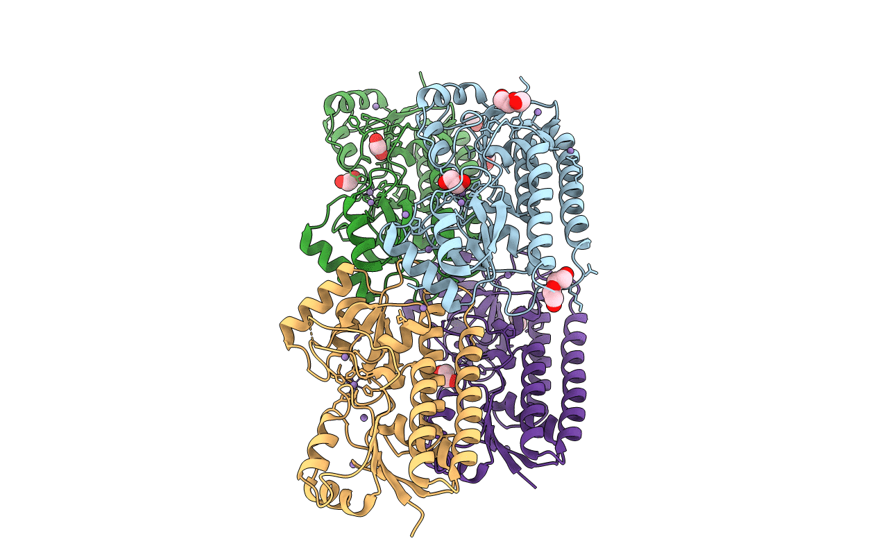
PDB ID:
6RU4
Keywords:
Title:
Structure of the SBP FpvC from pseudomonas aeruginosa in complex with Mn2+
Biological Source:
Source Organism(s):
Pseudomonas aeruginosa PAO1 (Taxon ID: 208964)
Expression System(s):
Method Details:
Experimental Method:
Resolution:
2.49 Å
R-Value Free:
0.23
R-Value Work:
0.19
R-Value Observed:
0.19
Space Group:
P 1 21 1
