6RTD image
Entry Detail
PDB ID:
6RTD
Keywords:
Title:
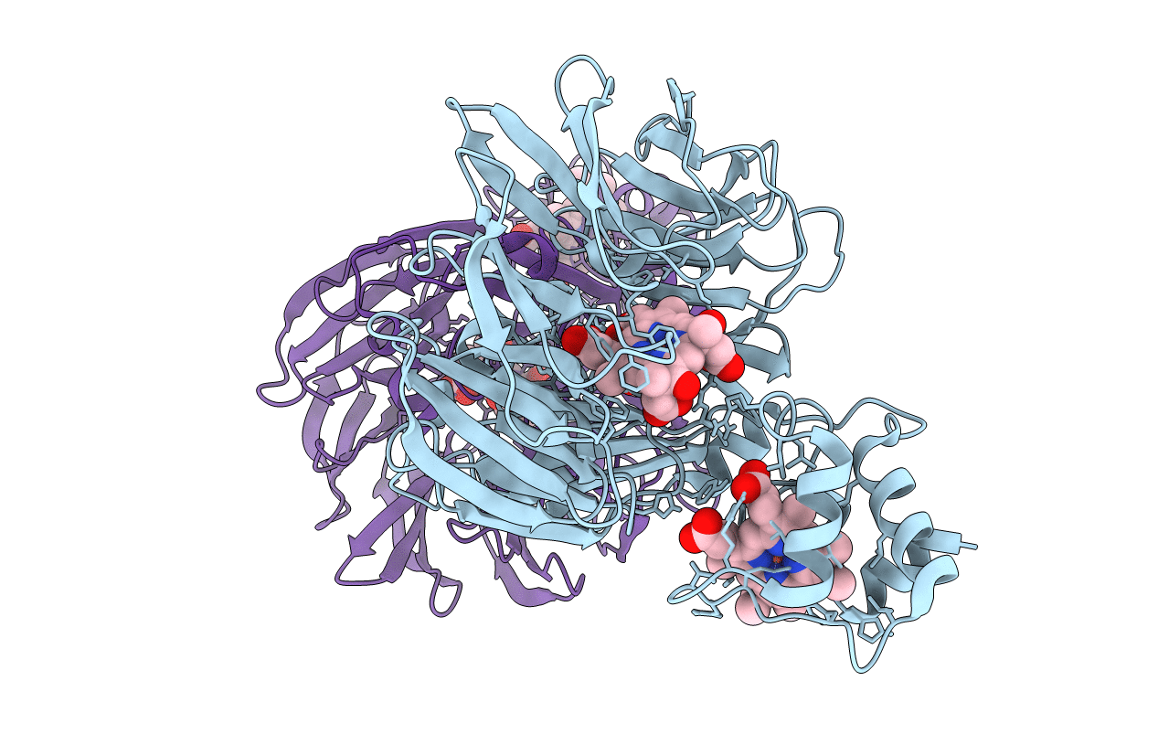
Dihydro-heme d1 dehydrogenase NirN in complex with DHE
Biological Source:
Source Organism:
Host Organism:
PDB Version:
Deposition Date:
2019-05-23
Release Date:
2019-06-19
Method Details:
Experimental Method:
Resolution:
2.36 Å
R-Value Free:
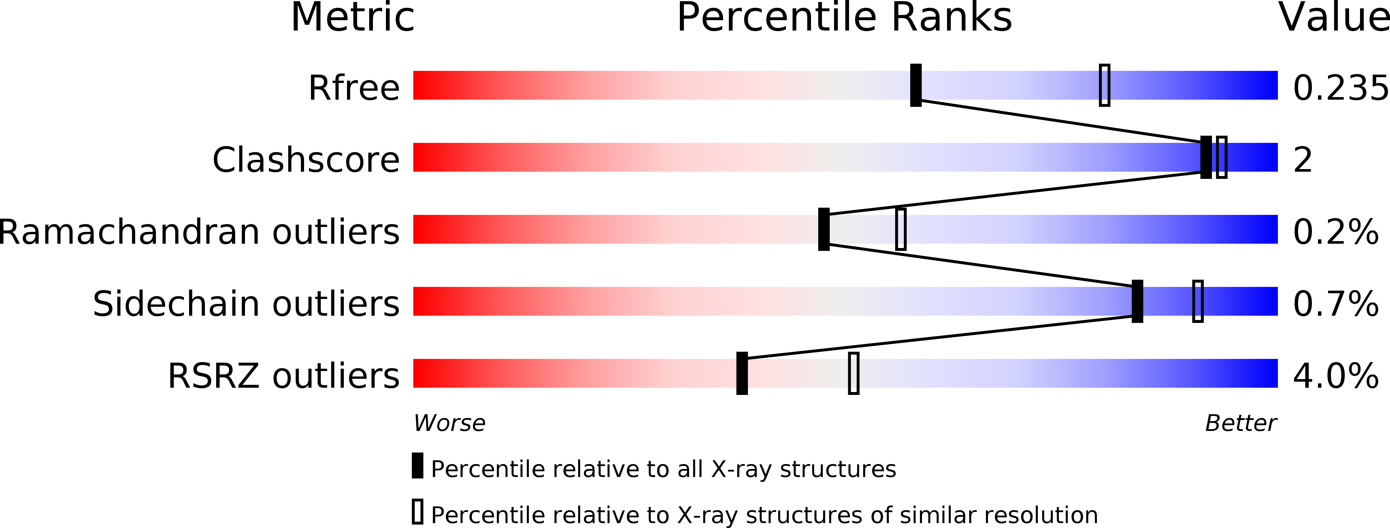
0.23
R-Value Work:
0.19
R-Value Observed:
0.19
Space Group:
C 1 2 1
Macromolecular Entities
Polymer Type:polypeptide(L)
Description:Cytochrome c
Chain IDs:A, B
Chain Length:509
Number of Molecules:2
Biological Source:Pseudomonas aeruginosa
Primary Citation
Crystal Structure of Dihydro-Heme d1Dehydrogenase NirN from Pseudomonas aeruginosa Reveals Amino Acid Residues Essential for Catalysis.
J.Mol.Biol. 431 3246 3260 (2019)
PMID: 31173777 DOI: 10.1016/j.jmb.2019.05.046

Abstact

Many bacteria can switch from oxygen to nitrogen oxides, such as nitrate or nitrite, as terminal electron acceptors in their respiratory chain. This process is called "denitrification" and enables biofilm formation of the opportunistic human pathogen Pseudomonas aeruginosa, making it more resilient to antibiotics and highly adaptable to different habitats. The reduction of nitrite to nitric oxide is a crucial step during denitrification. It is catalyzed by the homodimeric cytochrome cd1 nitrite reductase (NirS), which utilizes the unique isobacteriochlorin heme d1 as its reaction center. Although the reaction mechanism of nitrite reduction is well understood, far less is known about the biosynthesis of heme d1. The last step of its biosynthesis introduces a double bond in a propionate group of the tetrapyrrole to form an acrylate group. This conversion is catalyzed by the dehydrogenase NirN via a unique reaction mechanism. To get a more detailed insight into this reaction, the crystal structures of NirN with and without bound substrate have been determined. Similar to the homodimeric NirS, the monomeric NirN consists of an eight-bladed heme d1-binding β-propeller and a cytochrome c domain, but their relative orientation differs with respect to NirS. His147 coordinates heme d1 at the proximal side, whereas His323, which belongs to a flexible loop, binds at the distal position. Tyr461 and His417 are located next to the hydrogen atoms removed during dehydrogenation, suggesting an important role in catalysis. Activity assays with NirN variants revealed the essentiality of His147, His323 and Tyr461, but not of His417.

Legend

Protein

Chemical

Disease

Primary Citation of related structures