6RRQ image
Deposition Date 2019-05-20
Release Date 2020-04-15
Last Version Date 2024-01-24
Entry Detail
PDB ID:
6RRQ
Keywords:
Title:
Crystal structure of tyrosinase PvdP from Pseudomonas aeruginosa bound to copper
Biological Source:
Source Organism:
Method Details:
Experimental Method:
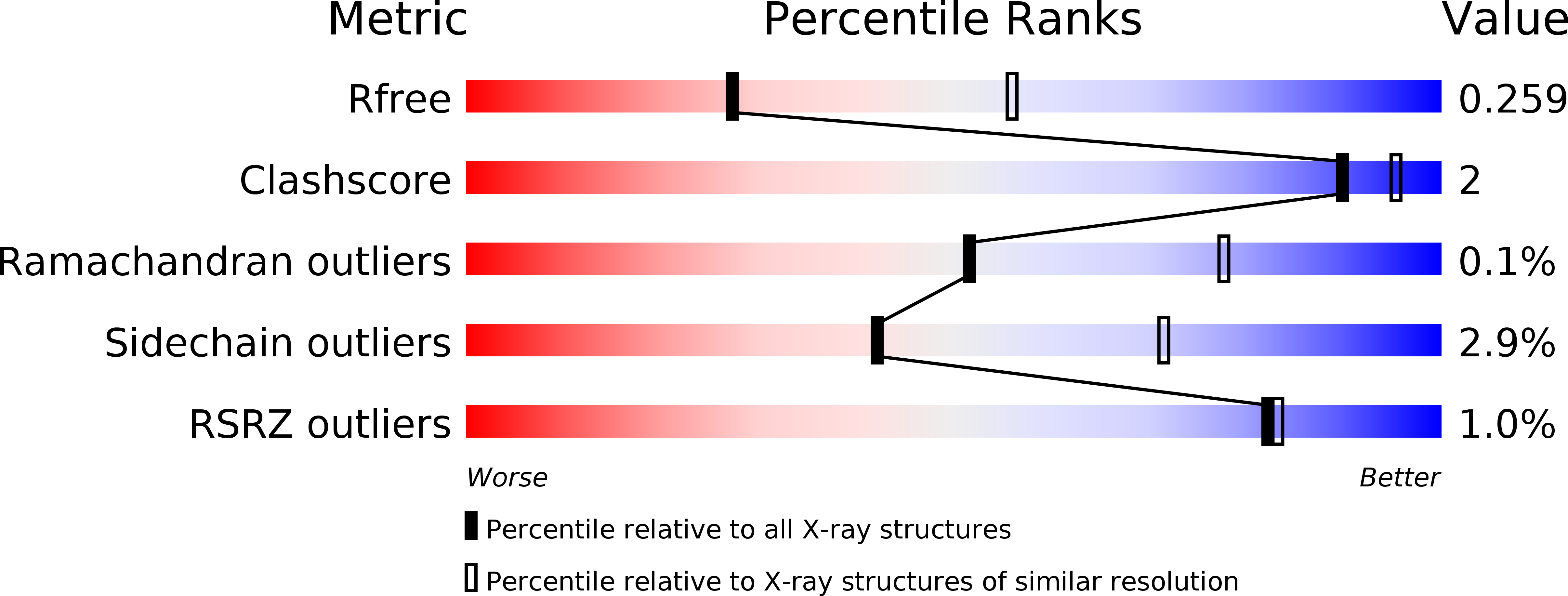
Resolution:
2.70 Å
R-Value Free:
0.25
R-Value Work:
0.18
R-Value Observed:
0.18
Space Group:
P 1 21 1
Macromolecular Entities
Polymer Type:polypeptide(L)
Molecule:PvdP
Gene (Uniprot):pvdP
Chain IDs:A, B
Chain Length:544
Number of Molecules:2
Biological Source:Pseudomonas aeruginosa PAO1
Primary Citation
A novel mechanism of inhibition by phenylthiourea on PvdP, a tyrosinase synthesizing pyoverdine of Pseudomonas aeruginosa.
Int.J.Biol.Macromol. 146 212 221 (2020)
PMID: 31899238 DOI: 10.1016/j.ijbiomac.2019.12.252

Abstact

The biosynthesis of pyoverdine, the major siderophore of Pseudomonas aeruginosa, is a well-organized process involving a discrete number of enzyme-catalyzed steps. The final step of this process involves the PvdP tyrosinase, which converts ferribactin into pyoverdine. Thus, inhibition of the PvdP tyrosinase activity provides an attractive strategy to interfere with siderophore synthesis to manage P. aeruginosa infections. Here, we report phenylthiourea as a non-competitive inhibitor of PvdP for which we solved a crystal structure in complex with PvdP. The crystal structure indicates that phenylthiourea binds to an allosteric binding site and thereby interferes with its tyrosinase activity. We further provide proofs that PvdP tyrosinase inhibition by phenylthiourea requires the C-terminal lid region. This provides opportunities to develop inhibitors that target the allosteric site, which seems to be confined to fluorescent pseudomonads, and not the tyrosinase active site. Furthermore, increases the chances to identify PvdP inhibitors that selectively interfere with siderophore synthesis.

Legend

Protein

Chemical

Disease

Primary Citation of related structures