6RQO image
Deposition Date 2019-05-16
Release Date 2019-07-17
Last Version Date 2024-11-20
Entry Detail
PDB ID:
6RQO
Title:
Steady-state-SMX activated state structure of bacteriorhodopsin
Biological Source:
Method Details:
Experimental Method:
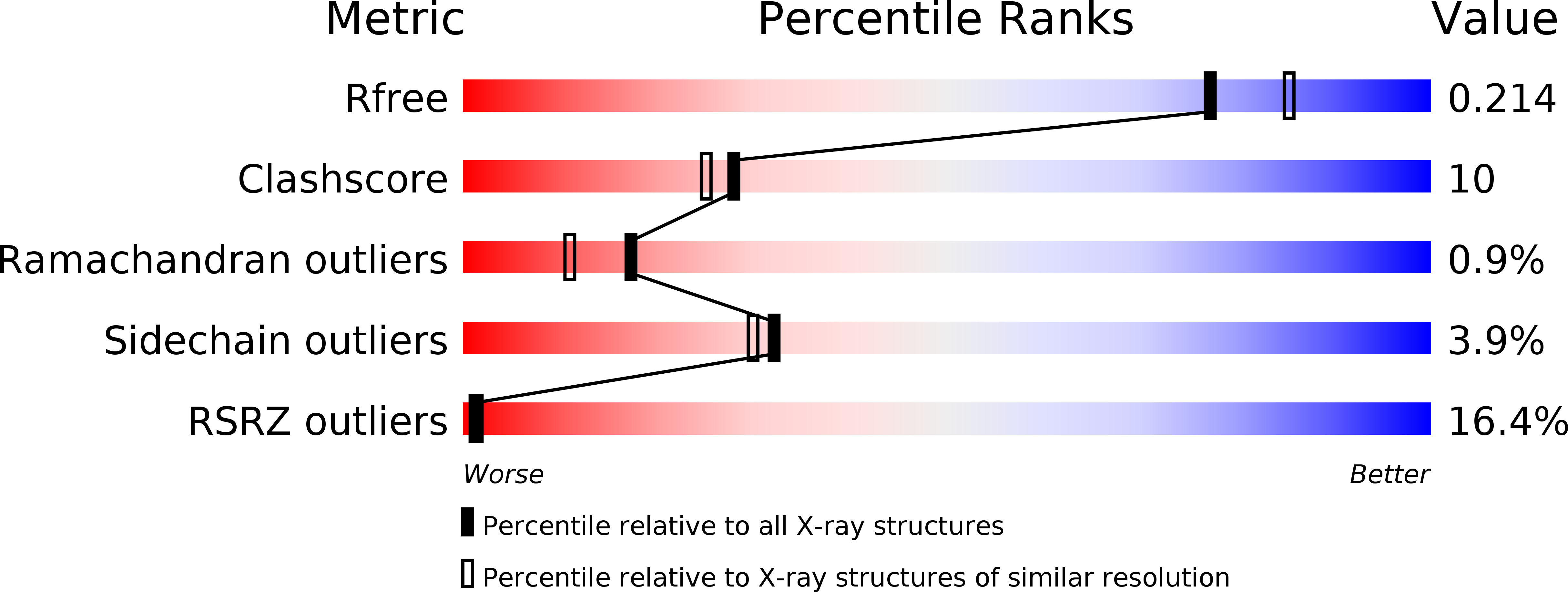
Resolution:
2.00 Å
R-Value Free:
0.24
R-Value Work:
0.20
R-Value Observed:
0.20
Space Group:
P 63
Macromolecular Entities
Structures with similar UniProt ID
Protein Blast
Polymer Type:polypeptide(L)
Molecule:Bacteriorhodopsin
Gene (Uniprot):bop
Chain IDs:A
Chain Length:223
Number of Molecules:1
Biological Source:Halobacterium salinarum (strain ATCC 700922 / JCM 11081 / NRC-1)
Ligand Molecules
Primary Citation
Proton uptake mechanism in bacteriorhodopsin captured by serial synchrotron crystallography.
Science 365 61 65 (2019)
PMID: 31273117 DOI: 10.1126/science.aaw8634

Abstact

Conformational dynamics are essential for proteins to function. We adapted time-resolved serial crystallography developed at x-ray lasers to visualize protein motions using synchrotrons. We recorded the structural changes in the light-driven proton-pump bacteriorhodopsin over 200 milliseconds in time. The snapshot from the first 5 milliseconds after photoactivation shows structural changes associated with proton release at a quality comparable to that of previous x-ray laser experiments. From 10 to 15 milliseconds onwards, we observe large additional structural rearrangements up to 9 angstroms on the cytoplasmic side. Rotation of leucine-93 and phenylalanine-219 opens a hydrophobic barrier, leading to the formation of a water chain connecting the intracellular aspartic acid-96 with the retinal Schiff base. The formation of this proton wire recharges the membrane pump with a proton for the next cycle.

Legend

Protein

Chemical

Disease

Primary Citation of related structures
Feedback Form
Name
Email
Institute
Feedback