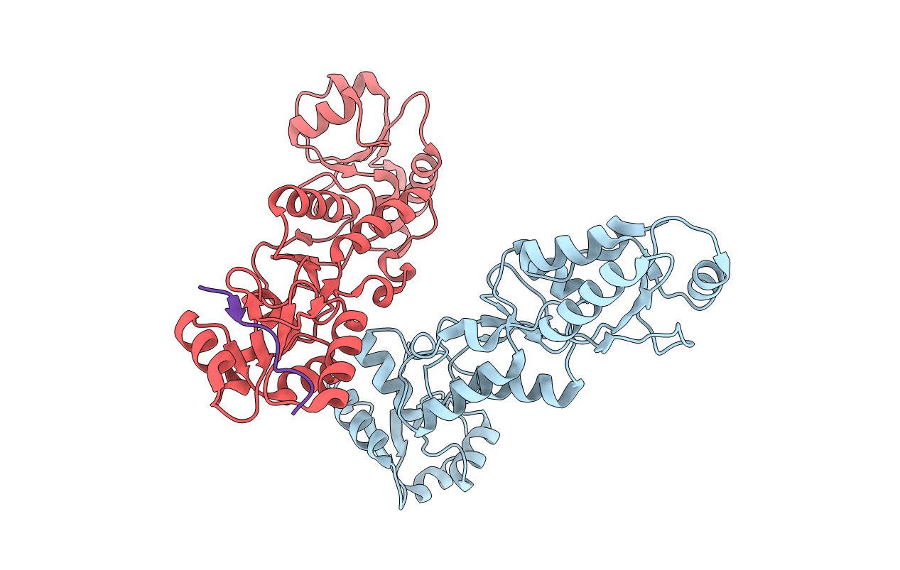
6RML image
Entry Detail
PDB ID:
6RML
Title:
Crystal structure of TOPBP1 BRCT0,1,2 in complex with a 53BP1 phosphopeptide
Biological Source:
Source Organism:
PDB Version:
Deposition Date:
2019-05-07
Release Date:
2019-06-12
Method Details:
Experimental Method:
Resolution:
2.81 Å
R-Value Free:
0.25
R-Value Work:
0.22
R-Value Observed:
0.22
Space Group:
P 32 2 1
Macromolecular Entities
Polymer Type:polypeptide(L)
Description:DNA topoisomerase 2-binding protein 1
Chain IDs:A (auth: B), B (auth: A)
Chain Length:292
Number of Molecules:2
Biological Source:Homo sapiens
Polymer Type:polypeptide(L)
Description:53BP1
Chain IDs:C
Chain Length:15
Number of Molecules:1
Biological Source:Homo sapiens
Ligand Molecules
Primary Citation
Phosphorylation-mediated interactions with TOPBP1 couple 53BP1 and 9-1-1 to control the G1 DNA damage checkpoint.
Elife 8 ? ? (2019)
PMID: 31135337 DOI: 10.7554/eLife.44353

Abstact

Coordination of the cellular response to DNA damage is organised by multi-domain 'scaffold' proteins, including 53BP1 and TOPBP1, which recognise post-translational modifications such as phosphorylation, methylation and ubiquitylation on other proteins, and are themselves carriers of such regulatory signals. Here we show that the DNA damage checkpoint regulating S-phase entry is controlled by a phosphorylation-dependent interaction of 53BP1 and TOPBP1. BRCT domains of TOPBP1 selectively bind conserved phosphorylation sites in the N-terminus of 53BP1. Mutation of these sites does not affect formation of 53BP1 or ATM foci following DNA damage, but abolishes recruitment of TOPBP1, ATR and CHK1 to 53BP1 damage foci, abrogating cell cycle arrest and permitting progression into S-phase. TOPBP1 interaction with 53BP1 is structurally complimentary to its interaction with RAD9-RAD1-HUS1, allowing these damage recognition factors to bind simultaneously to the same TOPBP1 molecule and cooperate in ATR activation in the G1 DNA damage checkpoint.

Legend

Protein

Chemical

Disease

Primary Citation of related structures